


Chapter 1 of 5 · Surgical Management
Port placement
Port placement and Percuvance instrument insertion technique
Expert statements on this page
No expert statements were drawn from this page.
Host summaries · secondary, not cited in answers
Educational content from recorded physician discussions — not medical advice. Talk to your (or your child's) care team about your situation.
Video
Lap Nissen trinity narrated
Dr. Steve Rothenberg · 5 min · Published May 2026
Video
Esophageal Disconnect for Severe GERD in Neurologically-Impaired Children:...
34 min · Published Aug 2017
Video
Gastroesophageal Reflux: Contemporary Management Pediatric Surgery Update...
32 min · Published Jul 2017
Video
QUAD #10 Pt.2: Surgical Technique for Thoracoscopic Repair of Tracheoesophageal Fistula (TEF) with Dr. Steve Rothenberg
Dr. Steve Rothenberg · Published Jun 2024
Video
22. HerniaTalk LIVE Q&A: Pediatric Hernias
Todd Ponsky · 59 min · Published Jul 2026
Podcast
Gastroesophageal Reflux Disease
Jeffrey Ponsky · 33 min · Published Jun 2016
Video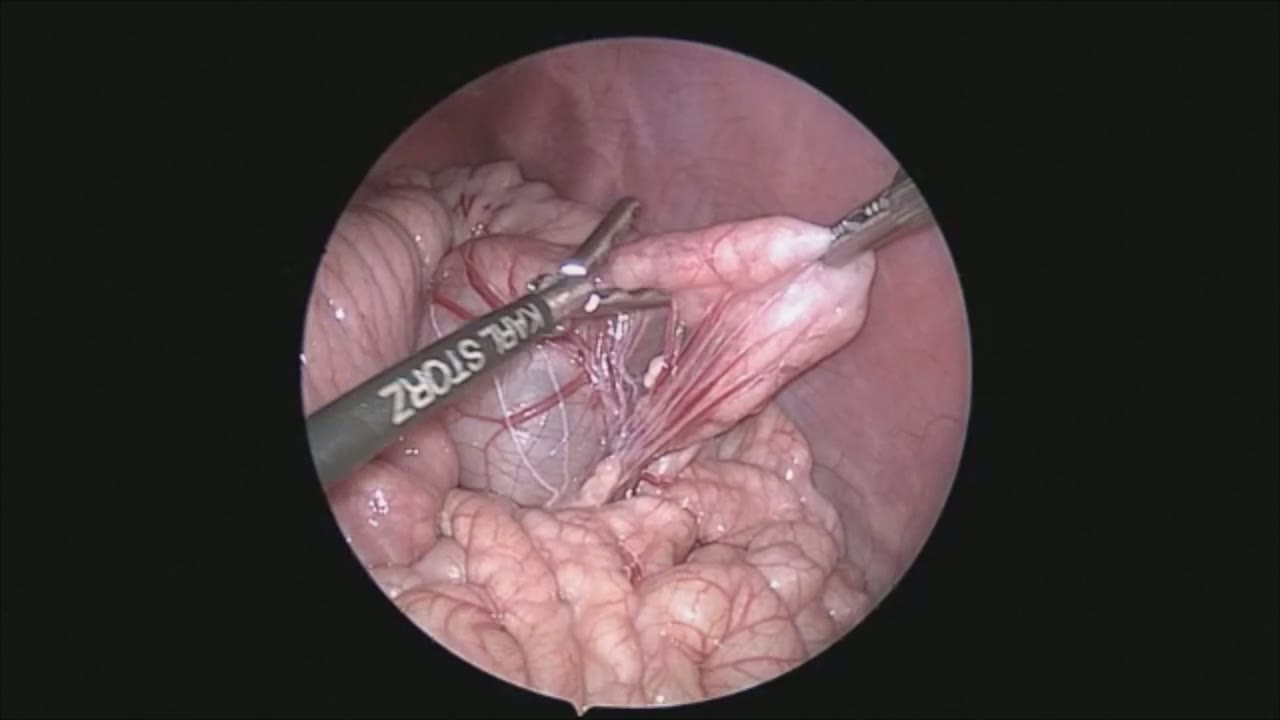
sealer demo
Dr. Steve Rothenberg · 3 min · Published May 2026
Video
pyloric knife
Dr. Steve Rothenberg · Published May 2026
Video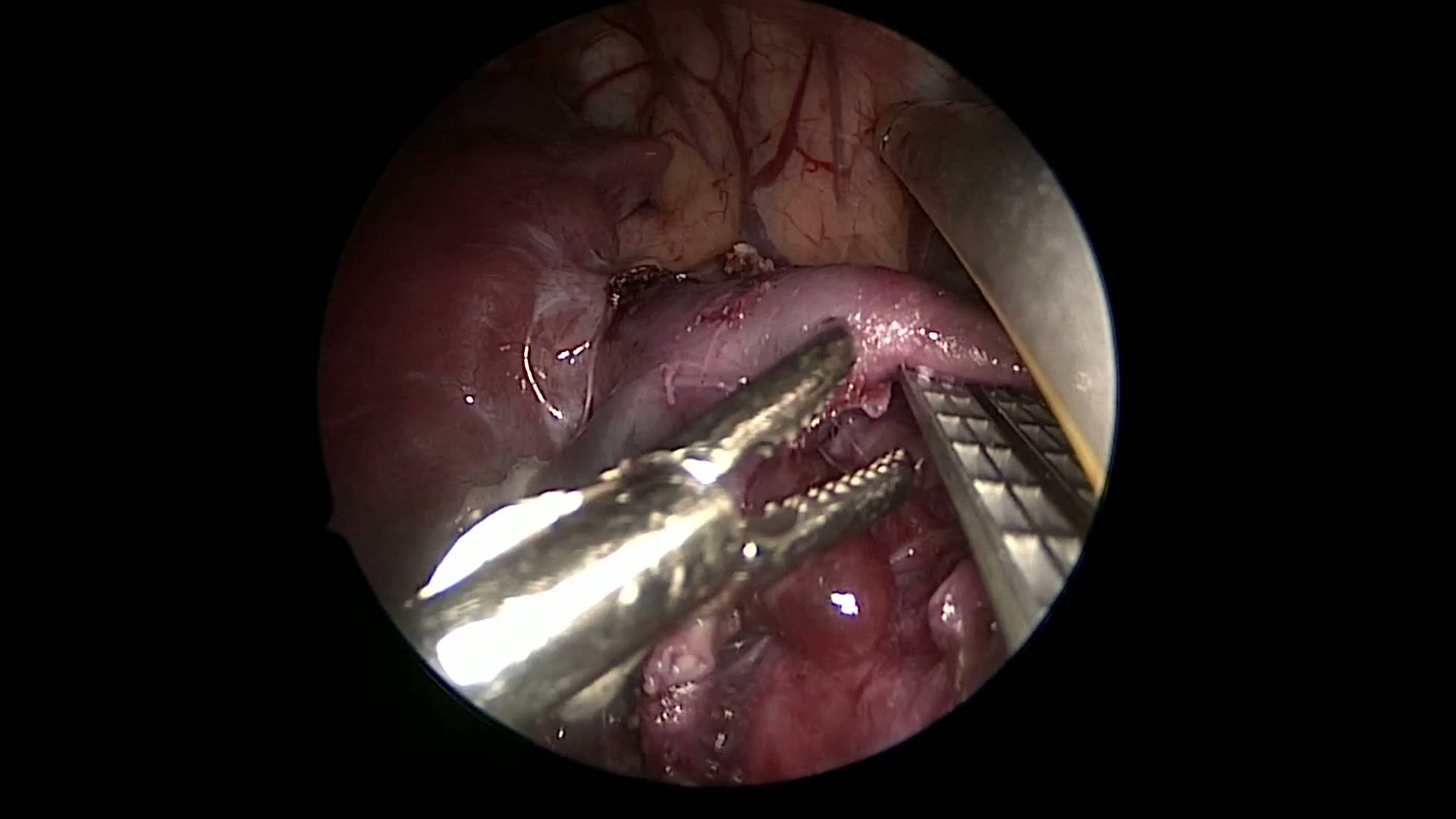
pulm artery LLL
Dr. Steve Rothenberg · 9 s · Published May 2026
Video
plication narrated
Dr. Steve Rothenberg · 6 min · Published May 2026
Video
open bowel anast
Dr. Steve Rothenberg · Published May 2026
Video
open bowel anast stapled
Dr. Steve Rothenberg · Published May 2026
Video
Intestinal Atresia Types Explained: Grossfeld Classification for Pediatric Surgery
1 min · Published Sep 2026
Video
Derivation and validation of the Pediatric Community-Acquired Pneumonia Severity (PedCAPS) score: A prospective cohort study
50 s · Published Sep 2026
Video
FETO for Late-Diagnosed Severe Congenital Diaphragmatic Hernia (CDH) at Cincinnati Children's with Dr. Beth Rymeski
4 min · Published Sep 2026
Video
Severe Congenital Diaphragmatic Hernia (CDH) Case: FETO Management & Unexpected Findings with Dr. Beth Rymeski
3 min · Published Sep 2026
Video
Fetoscopic Endoluminal Tracheal Occlusion (FETO) Procedure: Step-by-Step Guide with Dr. Beth Rymeski
4 min · Published Sep 2026
Video
Choledochal Cyst Types Explained: Pediatric Surgery Fundamentals
1 min · Published Sep 2026
What the experts said
The case is a redo fundoplication in a 30 kg child using the 3 mm Percuvance system
The Percuvance instrument is inserted by first inserting a needle tip on the shaft, then inserting through a 5 mm trocar placed in the left mid quadrant, then attaching the grasper
Three Percuvance instruments are used, supplying both liver retraction and stomach manipulation
The stomach retractor needle tip is inserted directly through the abdominal wall and then a grasper is attached to retract on the stomach
The 5 mm effector tips allow for atraumatic manipulation of the stomach in this larger child
The previous wrap appears to be intact but is loosened, and there is a small hiatal hernia
Complete takedown of the wrap and exposure of the left crus is a key maneuver in redo fundoplication
A two-stitch crural repair was performed using 2-0 Ethibond suture
It is extremely important in redo fundoplication to completely take down the previous wrap so that the hiatal defect can be well visualized and repaired appropriately
The stomach is retracted down to further lengthen the esophagus to ensure there is adequate length
The wrap is formed going from stomach to the anterior wall of the esophagus, with the top stitch also including the anterior diaphragmatic rim
Including the anterior diaphragmatic rim in the top stitch helps lock the wrap into the abdomen and hopefully will prevent recurrent herniation
Three sutures are placed, each incorporating a bite of the stomach, a bite of the esophagus, and a bite of the wrap portion of the stomach
Removal of the three Percuvance tips took approximately one minute