


From
StayCurrentMD
Trinity Ileal resection w narration
With Dr. Steve Rothenberg
Part of
Crohn's Disease 11 items
Chapter 1 of 4 · Case-Based Learning
Case presentation
Case presentation and patient positioning
Expert statements on this page
No expert statements were drawn from this page.
Host summaries · secondary, not cited in answers
Educational content from recorded physician discussions — not medical advice. Talk to your (or your child's) care team about your situation.
Video
Journal of Pediatric Surgery Article Review: September 2021
Todd Ponsky · 11 min · Published Jul 2026
Video
Best of the Best Gen Surg - Therapeutic Effect of Oridonin against Inflammatory Bowel Disease Involves Inhibition of Intestinal Fibrosis via NF-kB and p38STAT1 Pathways - Dr. Mao
11 min · Published Sep 2022
Video
Inflammatory Bowel Disease (IBD) - Samir Pandya: Update Course 2014
28 min · Published Nov 2018
Podcast
Update Course Rewind: 2020 ERAS
11 min · Published Feb 2021
Video
Inflammatory Bowel Disease: Update Course 2014
28 min · Published Sep 2014
Video
ERAS - Clinical Practice Updates
Published Sep 2020
Video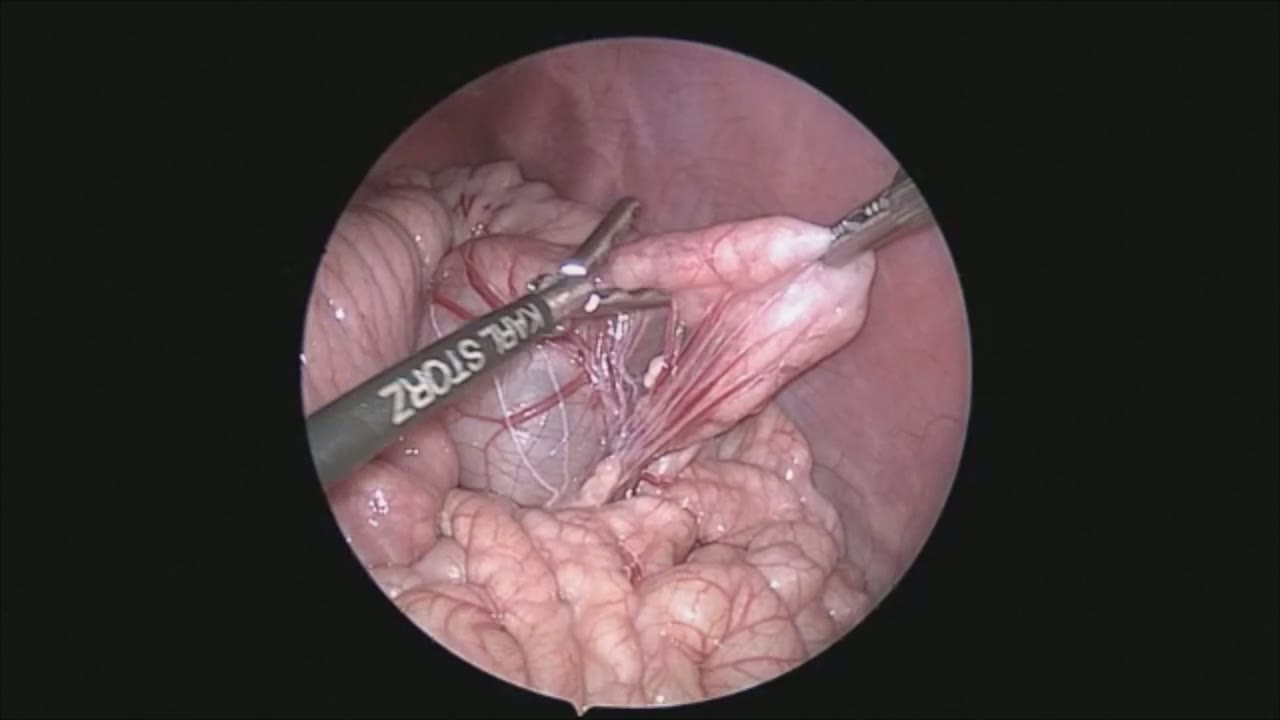
sealer demo
Dr. Steve Rothenberg · 3 min · Published May 2026
Video
pyloric knife
Dr. Steve Rothenberg · Published May 2026
Video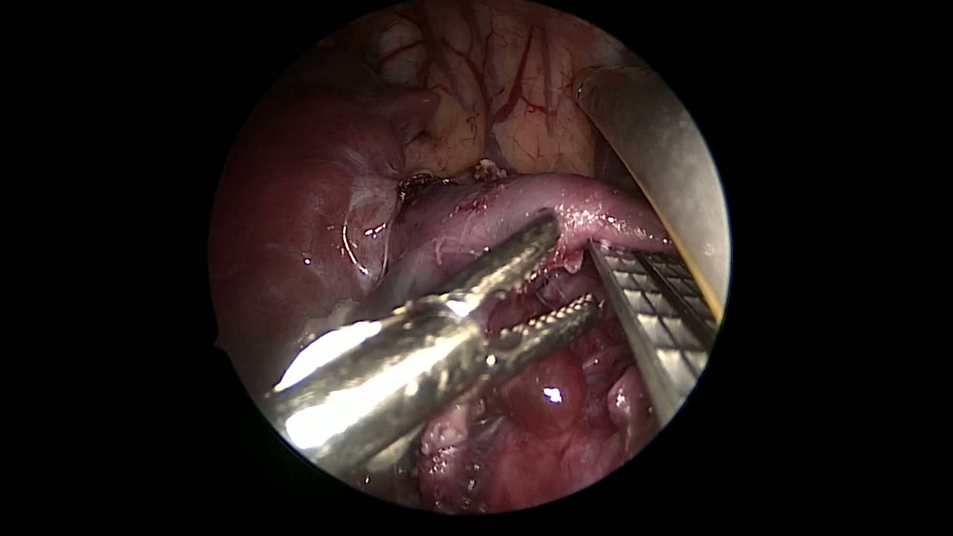
pulm artery LLL
Dr. Steve Rothenberg · 9 s · Published May 2026
Video
plication narrated
Dr. Steve Rothenberg · 6 min · Published May 2026
Video
percuvance redo nissen
Dr. Steve Rothenberg · 5 min · Published May 2026
Video
open bowel anast
Dr. Steve Rothenberg · Published May 2026
Video
Intestinal Atresia Types Explained: Grossfeld Classification for Pediatric Surgery
1 min · Published Sep 2026
Video
Derivation and validation of the Pediatric Community-Acquired Pneumonia Severity (PedCAPS) score: A prospective cohort study
50 s · Published Sep 2026
Video
FETO for Late-Diagnosed Severe Congenital Diaphragmatic Hernia (CDH) at Cincinnati Children's with Dr. Beth Rymeski
4 min · Published Sep 2026
Video
Severe Congenital Diaphragmatic Hernia (CDH) Case: FETO Management & Unexpected Findings with Dr. Beth Rymeski
3 min · Published Sep 2026
Video
Fetoscopic Endoluminal Tracheal Occlusion (FETO) Procedure: Step-by-Step Guide with Dr. Beth Rymeski
4 min · Published Sep 2026
Video
Choledochal Cyst Types Explained: Pediatric Surgery Fundamentals
1 min · Published Sep 2026
What the experts said
A 19 year old male presented with a 3 year history of medically managed Crohn's disease with increasing abdominal pain and intermittent vomiting
MRI showed a fixed stricture of the terminal ileum with gross proximal dilatation
There was approximately a 10 centimeter length of ileum proximal to the ileocecal valve which remained pliable and not grossly involved
The decision was made to preserve the ileocecal valve based on finding 10 cm of relatively uninvolved distal ileum
The 5 millimeter Cool Seal Trinity device can be used to dissect through thick mesentery and create mesenteric windows
When mesentery is very thick, it can be taken in two separate leaflets (one proximal and one distal) while maintaining complete hemostasis
A useful technique for thick mesentery is to make a proximal seal on the mesenteric base and then a more distal seal near the bowel wall, dividing tissue at this level to ensure no incomplete seal and maintain proximal vascular control
The mesentery was almost 2 centimeters thick in this case
By using the 5 mm device for mesenteric division, trocars could be limited to two 5 millimeter ports and one 12 millimeter port, which is cosmetically superior to requiring larger ports for 10 mm sealer or 12 mm stapler
The mesenteric division portion of the operation took approximately 20 minutes with no significant blood loss and no unsealed vessels
The patient weighs approximately 62 kg
EndoGIA tan loads were used to divide the bowel both proximally and distally
The 5 mm device has no heat spread and is immediately cool, allowing manipulation of bowel and surrounding structures using the grasper function without needing to exchange for a different grasping tool
Two loads of the endoGIA were required to completely divide the grossly dilated proximal bowel
A 60 millimeter load of the endoGIA was used to perform the side-to-side anastomosis
The resultant enterotomy was closed with a running 2-0 Vicryl suture
The operation took approximately 90 minutes
The patient was left with an NG tube overnight which was removed the next morning
The patient was started on a clear liquid diet on the 2nd postoperative day
The patient was discharged to home on the 3rd postoperative day
There was no blood loss during the procedure