


From
StayCurrentMD
Narrated R Adrenal Coolseal
With Dr. Steve Rothenberg
Part of
Pediatric Oncology 696 items
Chapter 1 of 4 · Case-Based Learning
Case presentation
Case presentation and initial mobilization
Expert statements on this page
No expert statements were drawn from this page.
Host summaries · secondary, not cited in answers
Educational content from recorded physician discussions — not medical advice. Talk to your (or your child's) care team about your situation.
Video
2025 Pediatric Surgery Update Course - Updates in Oncology
21 min · Published Aug 2025
Video
Social Determinants of Health Influence on Survival in Wilms Tumor, Neuroblastoma, and Hepatoblastoma
1 min · Published Jan 2026
Video
Update Course Rewind: Staging and Management of Neuroblastoma 2022
Published Jun 2023
Podcast
Journal of Pediatric Surgery Article Review: The Disruption Score
12 min · Published Aug 2021
Video
The International Neuroblastoma Risk Group (INRG) staging system: an INRG Task Force report
59 s · Published Mar 2026
Video
Image-guided core-needle or surgical biopsy for neuroblastoma diagnosis in children
54 s · Published Feb 2024
Video
Oncology Tumor Board Showdown: Pediatric Oncology 5 Question Quiz
1 min · Published Oct 2026
Video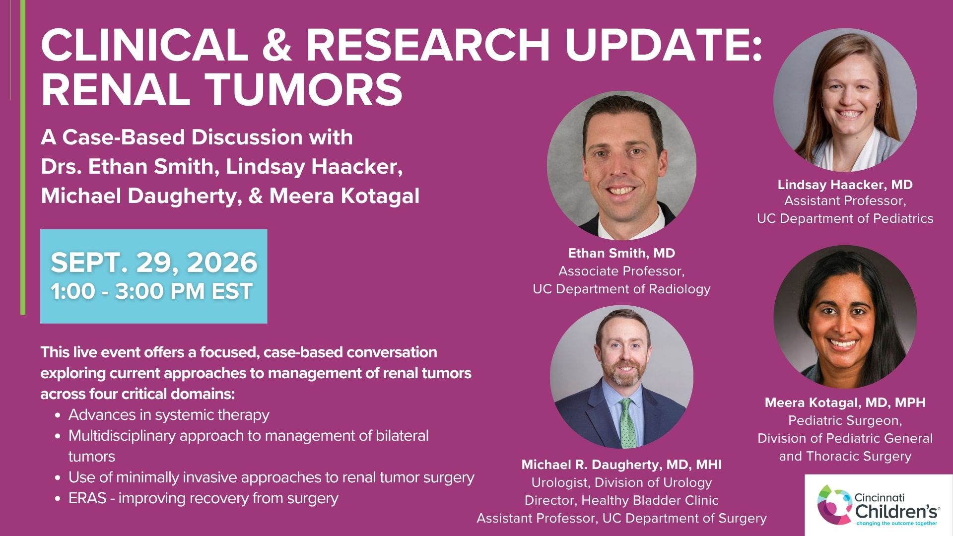
Clinical & Research Update: Renal Tumors with Drs. Ethan Smith, Lindsay Haacker, Michael Daugherty, and Meera Kotagal
71 min · Published Sep 2026
Video
Clinical & Research Update: Neuroblastoma with Drs. Katherine Somers, Cara Morin, Juan Gurria, and Meera Kotagal
67 min · Published Sep 2026
Video
Clinical & Research Update: Sarcoma w/ Drs. Roshni Dasgupta, Joseph Pressey, Arthur Meyer, Luke Pater
66 min · Published Sep 2026
Video
Update Course Rewind 2025: Nerve Monitoring in Pediatric Thyroid Surgery
6 min · Published Jul 2026
Video
Journal of Pediatric Surgery Article Review: June 2023, AAP Issue
Todd Ponsky · 13 min · Published Jul 2026
Video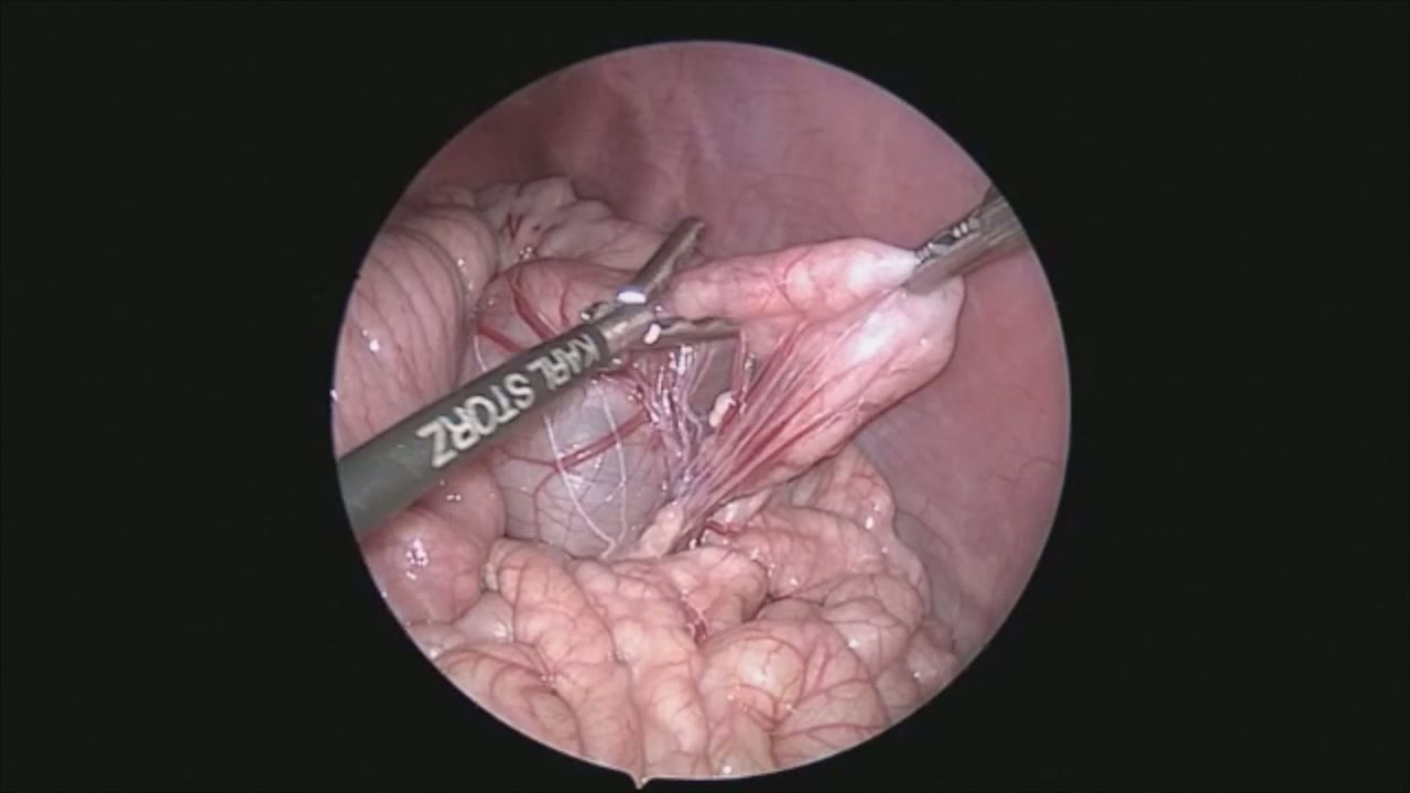
sealer demo
Dr. Steve Rothenberg · 3 min · Published May 2026
Video
pyloric knife
Dr. Steve Rothenberg · Published May 2026
Video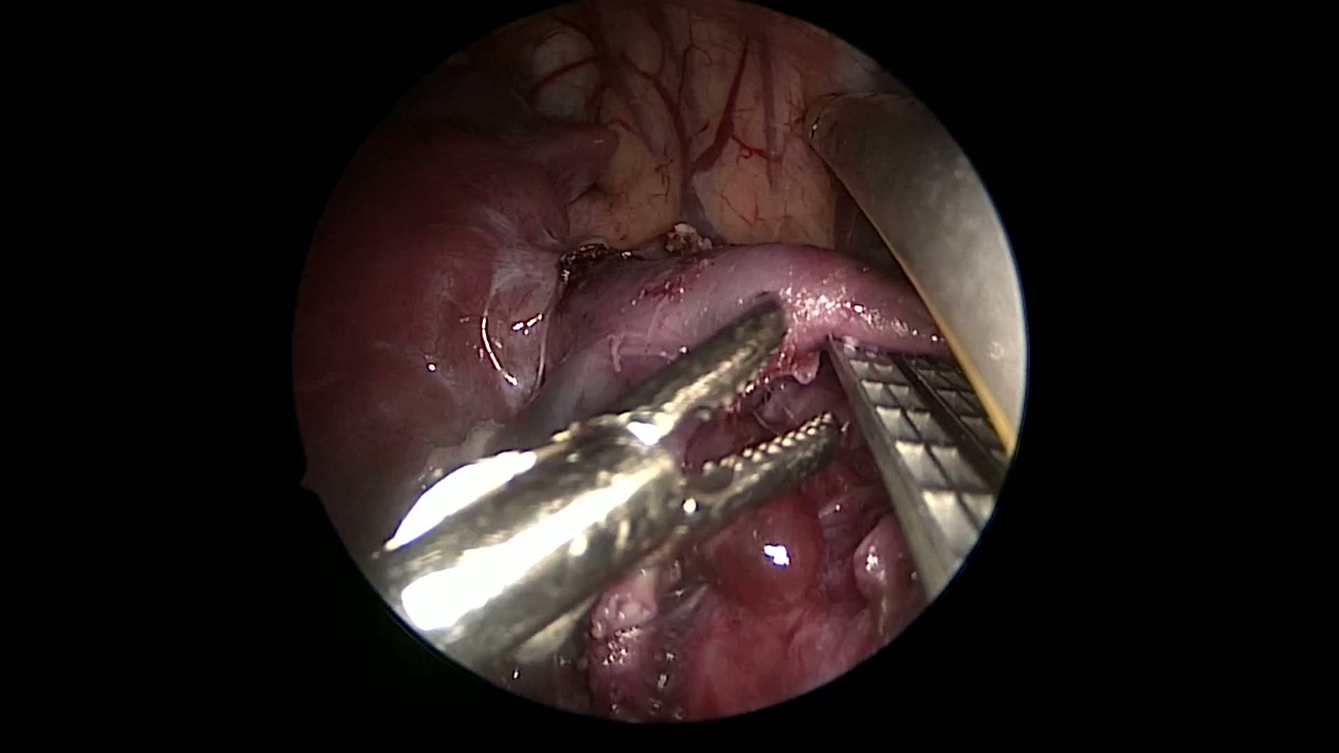
pulm artery LLL
Dr. Steve Rothenberg · 9 s · Published May 2026
Video
plication narrated
Dr. Steve Rothenberg · 6 min · Published May 2026
Video
percuvance redo nissen
Dr. Steve Rothenberg · 5 min · Published May 2026
Video
open bowel anast
Dr. Steve Rothenberg · Published May 2026
Video
Intestinal Atresia Types Explained: Grossfeld Classification for Pediatric Surgery
1 min · Published Sep 2026
Video
Derivation and validation of the Pediatric Community-Acquired Pneumonia Severity (PedCAPS) score: A prospective cohort study
50 s · Published Sep 2026
Video
FETO for Late-Diagnosed Severe Congenital Diaphragmatic Hernia (CDH) at Cincinnati Children's with Dr. Beth Rymeski
4 min · Published Sep 2026
Video
Severe Congenital Diaphragmatic Hernia (CDH) Case: FETO Management & Unexpected Findings with Dr. Beth Rymeski
3 min · Published Sep 2026
Video
Fetoscopic Endoluminal Tracheal Occlusion (FETO) Procedure: Step-by-Step Guide with Dr. Beth Rymeski
4 min · Published Sep 2026
Video
Choledochal Cyst Types Explained: Pediatric Surgery Fundamentals
1 min · Published Sep 2026
What the experts said
Patient was a 3½-year-old female who presented with chronic cough and chest pain, with CT scan showing right perirenal mass
The Cool Trinity 5 mm device has very low energy profile and heat spread, allowing dissection without worry of injury to the colon
The fine tips of the 5 mm jaw allow for both grasping and fine dissection between tissue planes
The tumor was directly on the inferior vena cava and densely adherent, as represented in the CT scan
The visualization afforded by the dual action and fine tips is better than all other tissue sealing devices and is equivalent to that of a 5 mm Maryland
At no time during this procedure was it necessary to remove the Trinity and insert a different grasping or dissecting device because of its ability to perform both functions
The device allows sealing and dividing tissues with separate actions, which is critically important for ensuring adequate hemostasis before division
The tissue between tumor and IVC was quite thick and inflamed, making the plane difficult to identify
The minimal heat spread ensures no damage to adjacent structures such as kidney, liver, bowel, or vena cava
There is minimal sticking following the sealing of tissues with the device
The device is taken out periodically to clean the jaws to remove any small amount of char
This procedure took under 2 hours
The tumor was placed in an endoscopic bag and brought out through an elongated umbilical incision, saving the child significant morbidity from a large open procedure
The child was discharged on the 2nd postoperative day
The pathology came back consistent with a neuroblastoma arising from the right adrenal gland