


Chapter 1 of 5 · Case-Based Learning
Case introduction
Case presentation and patient positioning
Expert statements on this page
No expert statements were drawn from this page.
Host summaries · secondary, not cited in answers
Educational content from recorded physician discussions — not medical advice. Talk to your (or your child's) care team about your situation.
Video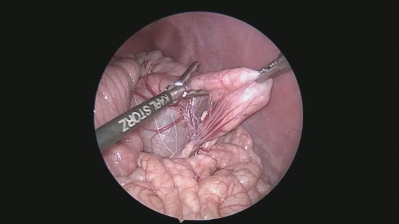
sealer demo
Dr. Steve Rothenberg · 3 min · Published May 2026
Video
pyloric knife
Dr. Steve Rothenberg · Published May 2026
Video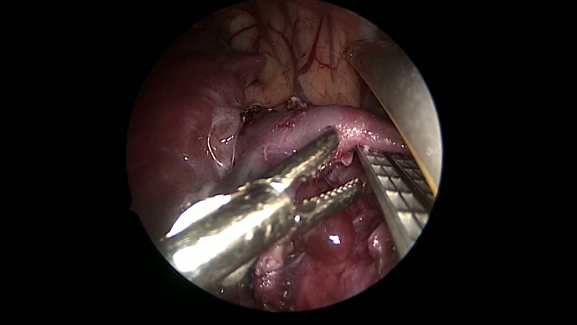
pulm artery LLL
Dr. Steve Rothenberg · 9 s · Published May 2026
Video
plication narrated
Dr. Steve Rothenberg · 6 min · Published May 2026
Video
percuvance redo nissen
Dr. Steve Rothenberg · 5 min · Published May 2026
Video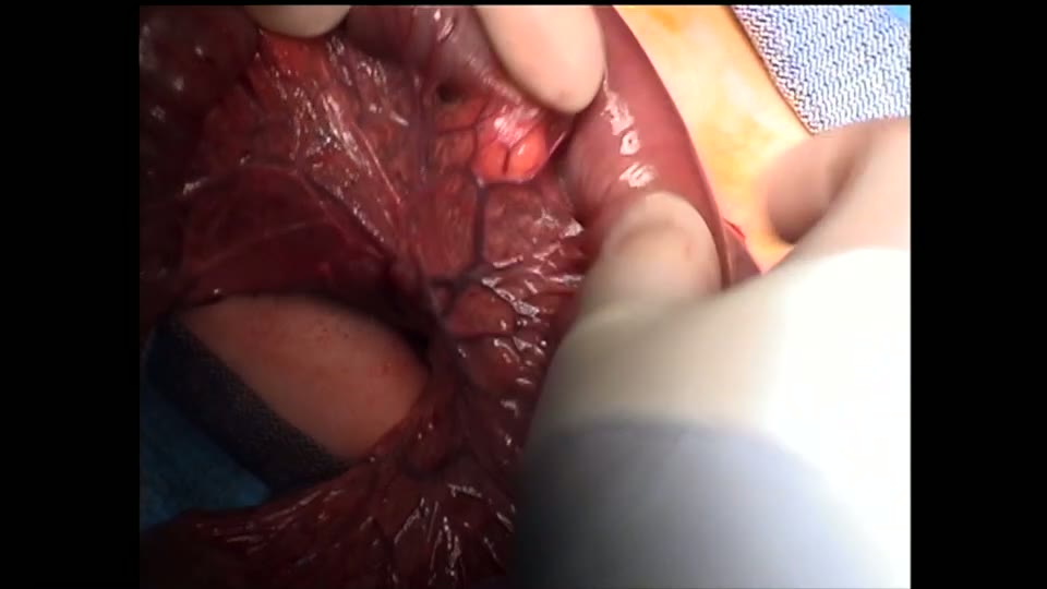
open bowel anast
Dr. Steve Rothenberg · Published May 2026
Video
Intestinal Atresia Types Explained: Grossfeld Classification for Pediatric Surgery
1 min · Published Sep 2026
Video
Derivation and validation of the Pediatric Community-Acquired Pneumonia Severity (PedCAPS) score: A prospective cohort study
50 s · Published Sep 2026
Video
FETO for Late-Diagnosed Severe Congenital Diaphragmatic Hernia (CDH) at Cincinnati Children's with Dr. Beth Rymeski
4 min · Published Sep 2026
Video
Severe Congenital Diaphragmatic Hernia (CDH) Case: FETO Management & Unexpected Findings with Dr. Beth Rymeski
3 min · Published Sep 2026
Video
Fetoscopic Endoluminal Tracheal Occlusion (FETO) Procedure: Step-by-Step Guide with Dr. Beth Rymeski
4 min · Published Sep 2026
Video
Choledochal Cyst Types Explained: Pediatric Surgery Fundamentals
1 min · Published Sep 2026
What the experts said
A 17 year old female with a 1 year history of chronic pain was found to have celiac artery compression and angulation on a CT angio
The procedure used 5 ports, 4 5-millimeter ports and 1 3-millimeter port
The ability to seal and cut safely with no heat spread is key for the entirety of the procedure
Identifying the right crus and where the right and left crus come together are critical for this procedure
There was unusually a large amount of inflammation in this area and a number of enlarged inflammatory nodes
Dissecting off inflamed nodes is critical in identifying the celiac trunk and to prevent inadvertent injury to the hepatic artery
The fibrous tissue in this area seemed to be impinging on the hepatic artery
A vessel loop was placed around the left gastric artery for traction and secured with 5 millimeter clips
It is necessary to divide celiac plexus nerve fibers to fully open up the area around the celiac trunk
All the energy is between the jaws and there is absolutely no heat spread outside of the jaws with the Trinity device
The lack of heat spread allows dissection, sealing, and dividing tissues while resting on the hepatic artery, celiac trunk, stomach, and other structures
The curved jaws allow better orientation to divide the fibers of the right crus and expose the median arcuate ligament
Two-hand dissection greatly enhances the surgery by allowing dissection, grasping tissue, and elevating planes with both hands
As the median arcuate ligament is divided, the celiac trunk expands and fills into view, showing significant compression of the structure
The ligament must be divided up to the insertion of the celiac trunk on the aorta to ensure complete division
The surgery lasted 90 minutes
The patient was discharged to home with complete resolution of her symptoms the following day