


From
StayCurrentMD
Lap Nissen trinity narrated
With Dr. Steve Rothenberg
Part of
Hiatal Hernia 3 items
Chapter 1 of 4 · Surgical Management
Setup & adhesiolysis
Patient positioning, trocar placement, and initial adhesiolysis
Expert statements on this page
No expert statements were drawn from this page.
Host summaries · secondary, not cited in answers
Educational content from recorded physician discussions — not medical advice. Talk to your (or your child's) care team about your situation.
Video
Esophageal Disconnect for Severe GERD in Neurologically-Impaired Children:...
34 min · Published Aug 2017
Video
QUAD #10 Pt.2: Surgical Technique for Thoracoscopic Repair of Tracheoesophageal Fistula (TEF) with Dr. Steve Rothenberg
Dr. Steve Rothenberg · Published Jun 2024
Video
22. HerniaTalk LIVE Q&A: Pediatric Hernias
Todd Ponsky · 59 min · Published Jul 2026
Video
Gastroesophageal Reflux: Contemporary Management Pediatric Surgery Update...
32 min · Published Jul 2017
Podcast
Gastroesophageal Reflux Disease
Jeffrey Ponsky · 33 min · Published Jun 2016
Podcast
Gastroesopheal Reflux Disease
33 min · Published Dec 2016
Video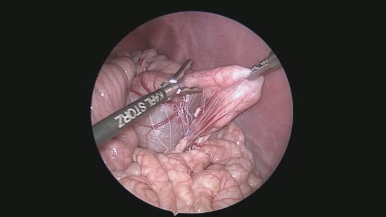
sealer demo
Dr. Steve Rothenberg · 3 min · Published May 2026
Video
pyloric knife
Dr. Steve Rothenberg · Published May 2026
Video
pulm artery LLL
Dr. Steve Rothenberg · 9 s · Published May 2026
Video
plication narrated
Dr. Steve Rothenberg · 6 min · Published May 2026
Video
percuvance redo nissen
Dr. Steve Rothenberg · 5 min · Published May 2026
Video
open bowel anast
Dr. Steve Rothenberg · Published May 2026
Video
Intestinal Atresia Types Explained: Grossfeld Classification for Pediatric Surgery
1 min · Published Sep 2026
Video
Derivation and validation of the Pediatric Community-Acquired Pneumonia Severity (PedCAPS) score: A prospective cohort study
50 s · Published Sep 2026
Video
FETO for Late-Diagnosed Severe Congenital Diaphragmatic Hernia (CDH) at Cincinnati Children's with Dr. Beth Rymeski
4 min · Published Sep 2026
Video
Severe Congenital Diaphragmatic Hernia (CDH) Case: FETO Management & Unexpected Findings with Dr. Beth Rymeski
3 min · Published Sep 2026
Video
Fetoscopic Endoluminal Tracheal Occlusion (FETO) Procedure: Step-by-Step Guide with Dr. Beth Rymeski
4 min · Published Sep 2026
Video
Choledochal Cyst Types Explained: Pediatric Surgery Fundamentals
1 min · Published Sep 2026
What the experts said
The Coseal Trinity 5mm device functions as both an excellent dissector with tips almost as fine as a Maryland and a vessel sealer.
Trocar placement includes 3 millimeter ports in the epigastrium for retraction and a 3 and 5 millimeter port in the left and right hand respectively for ancillary instruments.
The surgeon stands between the patient's legs with the cameraman on the surgeon's left.
Care is taken not to carry dissection high into the hiatus to avoid increased risk of hiatal hernia.
The anterior vagus nerve must be identified and preserved during retroesophageal dissection.
The gastrohepatic ligament is taken down to expose the right crus and create a safe retroesophageal window.
The Trinity device can seal and divide blood vessels up to 7 millimeters in size.
The Trinity device produces almost no heat spread, which prevents injury to the stomach, spleen, or surrounding organs.
The retroesophageal window is developed to eliminate the risk of injury to the esophagus or the posterior wall of the stomach.
The left crus is slightly cleaned to better allow for crural repair.
A small amount of peritoneum overlying the upper esophagus is taken to allow for proper placement in the wrap.
A 2-0 Ethibond suture on RB1 needle is used for crural repair in patients ranging from 2 kg to 100 kg.
Establishing good length of intraabdominal esophagus is key to developing a good wrap and preventing a slip Nissen.
The wrap should be tension-free and brought around behind the esophagus.
A small anterior bite of the esophagus is taken at the 10 and 11 o'clock position during wrap construction.
The stomach retractor is used to lengthen the esophagus by pulling downward, ensuring the wrap is above the GE junction.
Three stitches are used to complete the wrap.
The wrap is generally under 2 centimeters in length to ensure that an obstructive force is not created.
Great care is taken to make sure that the wrap is loose and there is no twisting of the esophagus.
The patient is started on clear liquids immediately after the procedure and discharged the following morning.