


Chapter 1 of 1 · Surgical Management
Plication technique
Laparoscopic Diaphragmatic Plication Technique
Expert statements on this page
No expert statements were drawn from this page.
Host summaries · secondary, not cited in answers
Educational content from recorded physician discussions — not medical advice. Talk to your (or your child's) care team about your situation.
Video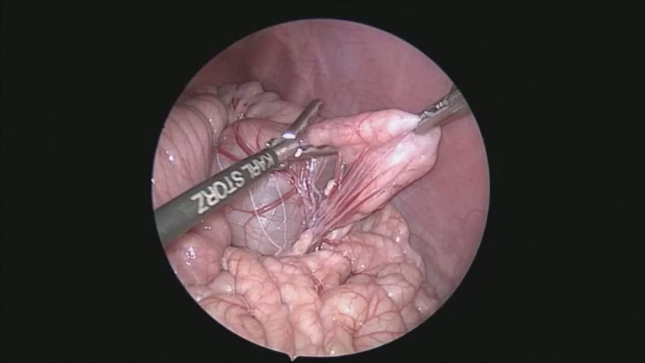
sealer demo
Dr. Steve Rothenberg · 3 min · Published May 2026
Video
pyloric knife
Dr. Steve Rothenberg · Published May 2026
Video
pulm artery LLL
Dr. Steve Rothenberg · 9 s · Published May 2026
Video
percuvance redo nissen
Dr. Steve Rothenberg · 5 min · Published May 2026
Video
open bowel anast
Dr. Steve Rothenberg · Published May 2026
Video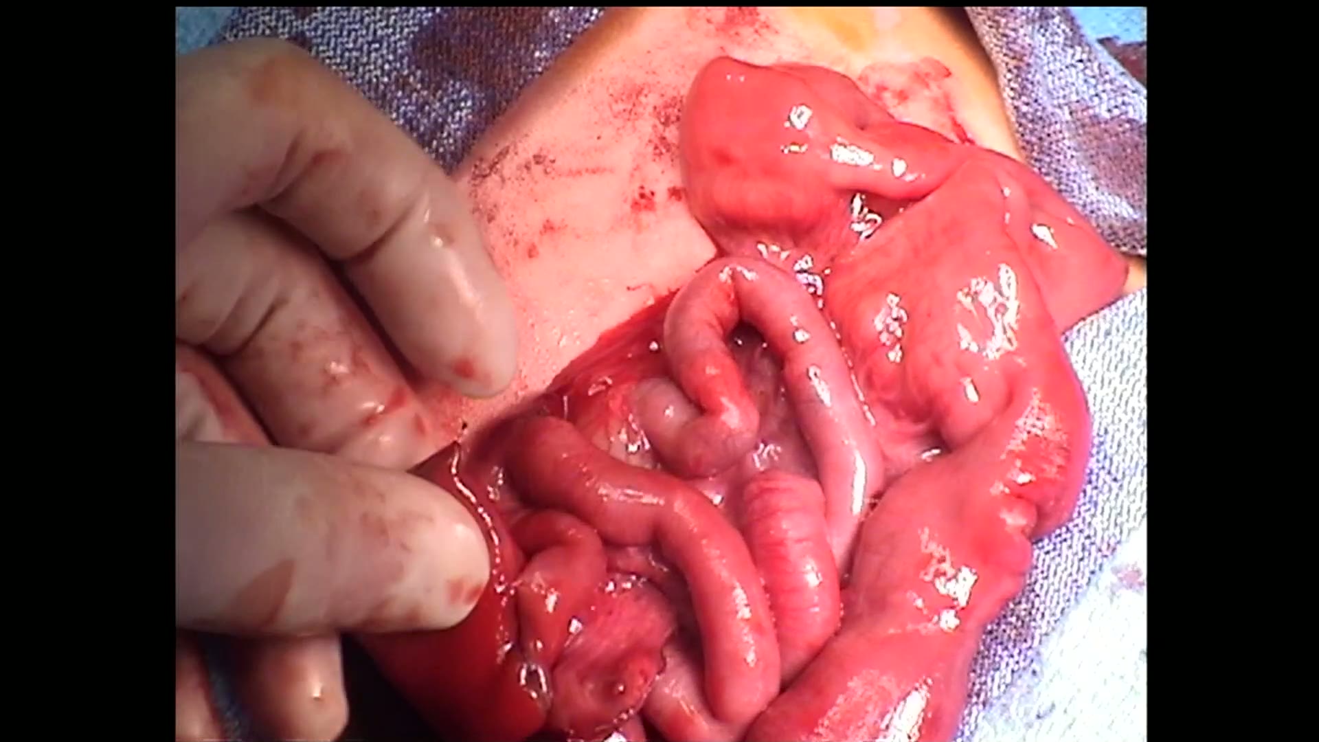
open bowel anast stapled
Dr. Steve Rothenberg · Published May 2026
Video
Intestinal Atresia Types Explained: Grossfeld Classification for Pediatric Surgery
1 min · Published Sep 2026
Video
Derivation and validation of the Pediatric Community-Acquired Pneumonia Severity (PedCAPS) score: A prospective cohort study
50 s · Published Sep 2026
Video
FETO for Late-Diagnosed Severe Congenital Diaphragmatic Hernia (CDH) at Cincinnati Children's with Dr. Beth Rymeski
4 min · Published Sep 2026
Video
Severe Congenital Diaphragmatic Hernia (CDH) Case: FETO Management & Unexpected Findings with Dr. Beth Rymeski
3 min · Published Sep 2026
Video
Fetoscopic Endoluminal Tracheal Occlusion (FETO) Procedure: Step-by-Step Guide with Dr. Beth Rymeski
4 min · Published Sep 2026
Video
Choledochal Cyst Types Explained: Pediatric Surgery Fundamentals
1 min · Published Sep 2026
What the experts said
Patient was a 3-month-old child with eventration of the right hemidiaphragm and significant respiratory issues.
The initial stitch is placed to take tension off the diaphragm; once surface tension is broken with the needle, 2 to 3 bites are performed to reef in the diaphragm.
A series of sutures are placed from medial to lateral to complete the first row of the plication.
Even in cases of right eventration, the diaphragm can easily be reached over the dome of the liver.
The speaker finds the transabdominal laparoscopic technique preferable to thoracoscopic plication because of greater access and mobility with trocars placed through the abdominal wall.
When performing thoracoscopic plication, trocars are fixed by the rib cage and motility and space are limited.
It is useful to tie the knots extracorporeally and use a knot pusher, allowing the assistant to hold down the liver with the left hand operating port while the knot pusher is being placed.
There is no risk of tearing these sutures out or causing other injury, so there is no reason to tie these sutures (which are under significant tension) intracorporeally.
Trocars are placed with the camera port in the umbilicus and right and left operating ports in the right and left mid quadrants.
When approaching the right hemidiaphragm, the left-sided port is placed slightly above the umbilicus to allow better angulation; the reverse is true for an elevated left hemidiaphragm.
A series of sutures are placed until adequate tension is felt to have been achieved on the diaphragm.
If one is not careful, the chest wall can be deformed in small infants because of the tension which can be placed on the diaphragm.
After the initial row of sutures is placed, it may be necessary to place a secondary row to achieve further plication.
In almost all cases, the speaker prefers to use a 2-0 Prolene suture on an RB-1 needle.