


Chapter 1 of 4 · Diagnosis & Workup
Diagnostic findings
Diagnostic laparoscopy findings for non-palpable undescended testicle
Expert statements on this page
No expert statements were drawn from this page.
Host summaries · secondary, not cited in answers
Educational content from recorded physician discussions — not medical advice. Talk to your (or your child's) care team about your situation.
Video
BOB Ped Surg 2023 - Jason Fraser, APSA - Presentation
Published Feb 2023
Video
Cryptorchidism Rapid Fire Session: Update Course 2015
6 min · Published Jul 2017
Video
22. HerniaTalk LIVE Q&A: Pediatric Hernias
Todd Ponsky · 59 min · Published Jul 2026
Video
Age at Orchiopexy JPS Review
Published Feb 2019
Video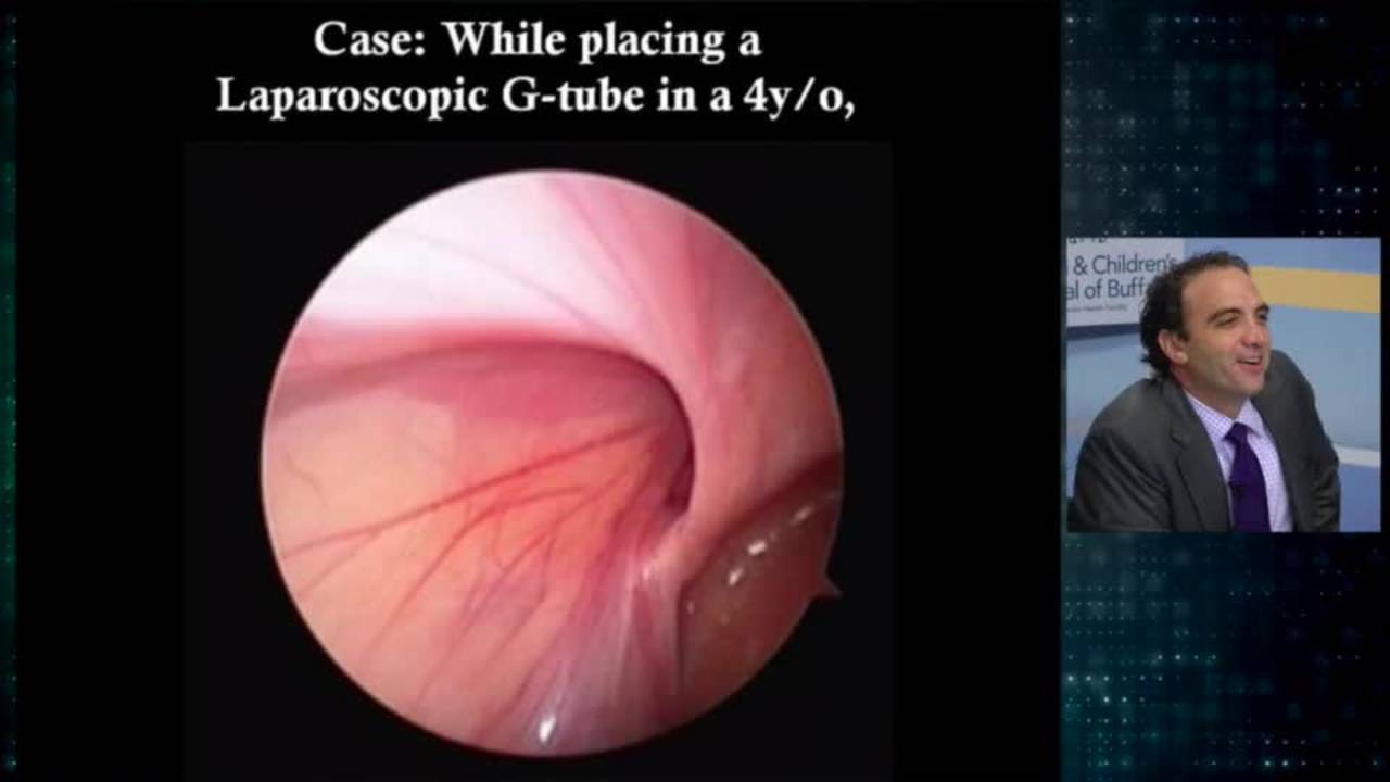
Groin Controversies: Update Course 2016
Dr. Todd Ponsky · 28 min · Published Jul 2017
Video
Pediatric Inguinal Hernia in Brief: Presentation, Workup, Diagnosis, Perioperative Considerations
Published Sep 2021
Video
Intestinal Atresia Types Explained: Grossfeld Classification for Pediatric Surgery
1 min · Published Sep 2026
Video
Derivation and validation of the Pediatric Community-Acquired Pneumonia Severity (PedCAPS) score: A prospective cohort study
50 s · Published Sep 2026
Video
FETO for Late-Diagnosed Severe Congenital Diaphragmatic Hernia (CDH) at Cincinnati Children's with Dr. Beth Rymeski
4 min · Published Sep 2026
Video
Severe Congenital Diaphragmatic Hernia (CDH) Case: FETO Management & Unexpected Findings with Dr. Beth Rymeski
3 min · Published Sep 2026
Video
Fetoscopic Endoluminal Tracheal Occlusion (FETO) Procedure: Step-by-Step Guide with Dr. Beth Rymeski
4 min · Published Sep 2026
Video
Choledochal Cyst Types Explained: Pediatric Surgery Fundamentals
1 min · Published Sep 2026
What the experts said
Laparoscopy is essential for the accurate diagnosis and optimal management of a non palpable undescended testicle.
When non-palpable undescended testicle is diagnosed, one of 3 possibilities will be confirmed on laparoscopy: an abdominal testicle, a blind ending vas (confirming absence of testicle), or vas deferens and testicular vessels exiting the deep ring (warranting inguinal exploration).
When an abdominal testicle is found, options include single-stage laparoscopic orchiopexy without vessel ligation, one or two-stage Fowler-Stevens procedure with vessel ligation, and the Shehata traction orchiopexy.
The procedure is performed using a single 5 millimeter trocar at the umbilicus for the camera and two lateral stab incisions on the right and left sides at or just below the level of the umbilicus for 3 millimeter instruments.
The mobilization of the testicle starts with stretching the gubernaculum and clearly visualizing the vas deferens, then the gubernaculum is divided as far away as possible from the vas to untether the testicle.
The vas should be kept in view at all times during gubernaculum division, and the surgeon should keep in mind the possibility of a long looping vas.
The vas is mobilized using blunt and sharp dissection with fine shears in a lateral to medial direction approaching the wall of the bladder.
The testicular vessels are mobilized by dividing the retroperitoneal attachments, which is where most of the length will be gained.
If the mobilized testicle is able to reach the contralateral ring, it is likely to reach the scrotum without further mobilization.
The point of entry for the needle-sheath trocar should be between the epigastric vessels and bladder edge, just over the pubic tubercle.
A full bladder actually aids in safe entry into the abdomen during trocar placement.
Following entry into the abdomen, a 10 millimeter trocar is introduced through the sheath to dilate the tunnel.
The testicle is grasped by the gubernaculum and brought through the trocar, taking care to keep it oriented correctly with the vas medial and vessels lateral.
The testicle is fixed in the dartos pouch using a slow absorbable suture.
It is not necessary to close the deep ring, as the incidence of inguinal hernia after laparoscopic orchiopexy is extremely low.
At 2 weeks postoperatively, tethering can be seen in the scrotum.
At 2 years postoperatively, the tethering is mostly resolved, and a testicle of normal caliber is easily visible and palpable in the right scrotum.