


From
StayCurrentMD
Anorectal Malformation Radiology: Pediatric Colorectal Controversies 2014
With Dr. Greg Bates
Chapter 1 of 8 · Diagnosis & Workup
Prenatal diagnosis
Prenatal diagnosis of anorectal malformations
Expert statements on this page
No expert statements were drawn from this page.
Host summaries · secondary, not cited in answers
Educational content from recorded physician discussions — not medical advice. Talk to your (or your child's) care team about your situation.
Podcast
Colorectal Quiz Episode 28: Female ARM Management - Perineal Fistula
19 min · Published Feb 2022
Video
ARMs in Neonates: Pediatric Colorectal Controversies 2014
105 min · Published Apr 2012
Video
Pediatric Colorectal Contraversies Part I: Pediatric Colorectal Contraversies...
Dr. Todd Ponsky · 25 min · Published Aug 2017
Video
Surgical Management Of Female Anorectal Malformation Patients Including...
57 min · Published Nov 2018
Video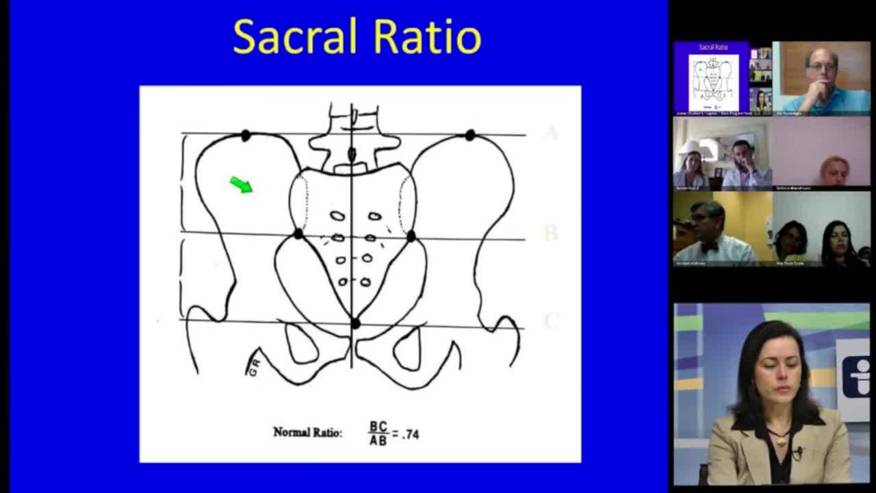
Panel Discussion and Case Presentation Part I: Pediatric Bowel Management 2013
Dr. Todd Ponsky · 12 min · Published May 2013
Podcast
Anorectal Malformations with Dr. Andrea Bischoff
47 min · Published Apr 2017
Video
Validation of an anorectal malformation trainer - Can a high-fidelity model simulate real life?
1 min · Published Jun 2026
Video
Complications and Long-Term Outcomes of Patients With Cloacal Malformation After Bowel Neovagina...
1 min · Published Jun 2026
Video
Safety and utility of long-acting steroid injection for management of post-operative stricture...
1 min · Published Jun 2026
Video
Association Between Social Determinants of Health and Choice of Urinary Reconstruction in Children
1 min · Published Jun 2026
Video
The Perineal Body Preserving PSARP (PPP)
11 min · Published Jun 2026
Video
Update Course Rewind 2025: Hirschsprung + ARM: Rare but Real
1 min · Published May 2026
Video
Intestinal Atresia Types Explained: Grossfeld Classification for Pediatric Surgery
1 min · Published Sep 2026
Video
Derivation and validation of the Pediatric Community-Acquired Pneumonia Severity (PedCAPS) score: A prospective cohort study
50 s · Published Sep 2026
Video
FETO for Late-Diagnosed Severe Congenital Diaphragmatic Hernia (CDH) at Cincinnati Children's with Dr. Beth Rymeski
4 min · Published Sep 2026
Video
Severe Congenital Diaphragmatic Hernia (CDH) Case: FETO Management & Unexpected Findings with Dr. Beth Rymeski
3 min · Published Sep 2026
Video
Fetoscopic Endoluminal Tracheal Occlusion (FETO) Procedure: Step-by-Step Guide with Dr. Beth Rymeski
4 min · Published Sep 2026
Video
Choledochal Cyst Types Explained: Pediatric Surgery Fundamentals
1 min · Published Sep 2026
What the experts said
Prenatal diagnosis of anorectal malformation in boys is not very good; it is often a surprise at birth.
In females, prenatal ultrasound showing bilateral hydronephrosis, small bladder, and dilated vagina suggests cloaca.
Fetal MRI is used for abnormal level 2 ultrasounds, especially with other anomalies, for prognostication and surgical planning.
Calcified enteroliths in the GI tract on neonatal KUB indicate urine mixing with meconium, suggesting a recto-GU fistula at least at the bulbar level or higher.
Patients with hydrocolpos can have significant urinary compromise with creatinine rising into the twos and threes if not managed urgently.
After 20 to 24 weeks gestation, T1 hyperintensity of meconium within the colon should be visible on fetal MRI; loss of T1 hyperintensity and bright T2 signal suggests urine mixing.
On fetal MRI, the distal rectum should extend at least 10 mm below the bladder neck in the second trimester, up to 20–24 mm in the third trimester; high position suggests ARM.
Hemisacrum (scimitar sacrum) is associated with presacral mass (teratoma or anterior myelomeningocele) and Currarino triad.
When hemisacrum is seen in a patient with anal malformation, Currarino triad must be assumed and presacral mass must be looked for.
For sacral ratio measurement, a true AP pelvis (not angled sacral view) and lateral pelvis are required; the lateral view is more accurate.
Normal sacral ratio ranges from 0.6 to 1.0; below 0.3 predicts very low likelihood of continence.
Sacral ratio measurement has inter-observer variability, especially in abnormal sacra, and is not as exact as often assumed.
Cross-table lateral (invertogram) must be performed after 24 to 36 hours to allow enough air and pressure to show the true level of the distal rectal pouch.
On invertogram, rectum within 1 cm of the perineal marker (BB) is considered safe for primary repair in selected cases.
Perineal ultrasound is more accurate than invertogram according to some radiologists, with ability to visualize fistulae and avoid radiation, but requires experience and standardized protocol.
Complications of colostomy double the incidence of complications in ARM patients, so avoiding colostomy when safe is desirable.
It is never wrong to perform a colostomy if uncertain about the level of the malformation.
In boys, approximately 95% have a fistula (outside of Down syndrome); the radiologist's job is to demonstrate it on distal colostography.
High-pressure distal colostography requires adequate pressurization to demonstrate fistulae; a small 'beak' off the rectal base indicates fistula location.
On distal colostography lateral view, the full sacrum should be included to show the relationship of the fistula to the tip of the sacrum.
It is difficult to distinguish radiologically between prostatic and bulbar urethral fistulae; the bend in the urethra is used as an approximate landmark.
Distal colonic segments that are very dilated and atonic are at higher risk of perforation during colostography due to Laplace's law (thin wall, high pressure).
Perforation during colostography with hyperosmotic contrast (e.g., cystoconray, ~400 mOsm) causes immediate fluid shift and peritonitis requiring emergent IV fluids and surgical consultation.
Iso-osmotic contrast is now preferred for distal colostography to avoid fluid shifts if perforation occurs.
Sacral ratio is one of four components of an ARM index (perineum appearance, malformation type, sacral index, tethered cord) being developed to predict continence and compare outcomes across institutions.