


From
Grand Rounds
Abdominal Trauma with Dr. Richard Falcone
With Dr. Rich Falcone
Part of
Blunt Abdominal Trauma 8 items
Chapter 1 of 6 · Fundamentals
CT imaging rationale
Introduction and rationale for selective CT imaging in pediatric blunt abdominal trauma
Expert statements on this page
No expert statements were drawn from this page.
Host summaries · secondary, not cited in answers
Educational content from recorded physician discussions — not medical advice. Talk to your (or your child's) care team about your situation.
Podcast
Pediatric Trauma With Dr. Richard Falcone
56 min · Published May 2017
Video
Guías APSA actualizadas para el tratamiento de lesiones contusas del hígado y del bazo
Published Nov 2023
Video
Update Course Rewind: Updates in Solid Organ Injury Management 2023
Published Mar 2024
Video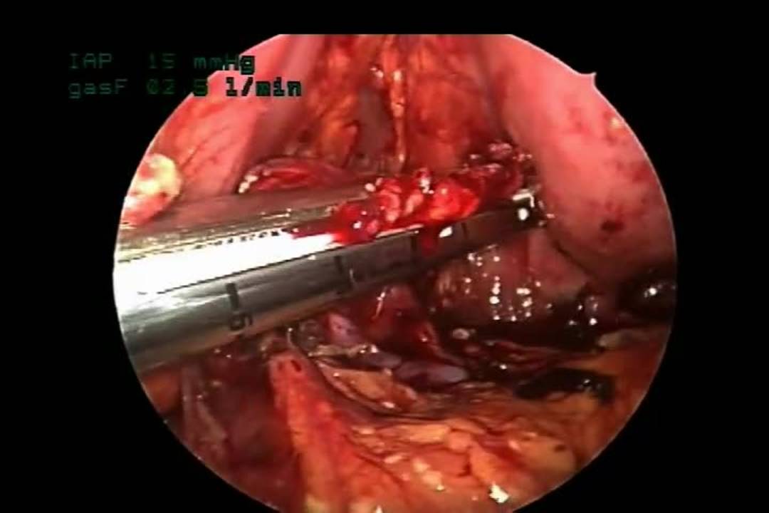
Laparoscopic Distal Pancreatectomy for Traumatic Transection
4 min · Published Nov 2019
Video
Trauma Talk Summary: Update Course 2017
Published Sep 2017
Video
Isolated low-grade solid organ injuries in children following blunt abdominal trauma
Published May 2023
Video
GYN #4 Management of Tubal Torsion: When to Consider Salpingectomy with Dr. Lesley Breech
CCHMC Pediatric Surgery · Published Oct 2024
Podcast
Pyloric Stenosis with Dr. Alex Bondoc
16 min · Published Jun 2024
Video
GYN #3 Management of an Adnexal Torsion with a Healthy Appearing Ovary with Dr. Lesley Breech
CCHMC Pediatric Surgery · Published May 2024
Video
GYN #2 Oophoropexy in Adnexal Torsion with Dr. Lesley Breech
CCHMC Pediatric Surgery · Published May 2024
Video
GYN #1 Importance of Documenting Reproductive Anatomy with Dr. Lesley Breech
CCHMC Pediatric Surgery · Published Feb 2024
Podcast
Esophageal Replacement with Dr. Dan von Allmen
13 min · Published Nov 2023
What the experts said
Six clinical factors (normal blood pressure for age, normal abdominal exam, AST less than 200, hematocrit greater than 30, and normal chest X-ray) have a 99% negative predictive value for intra-abdominal injury requiring CT scan.
Cincinnati Children's Hospital removed trauma labs or belly labs from initial lab draws approximately 8 years ago because they were wasting money and causing multiple IV attempts in children.
The only labs acutely needed for pediatric trauma are type and screen, CBC or gas, and coagulation studies for brain injury patients; belly labs should not be part of routine panel.
Dr. Falcone admitted a Grade 4 splenic injury to the floor rather than ICU based on clinical stability, despite institutional protocol concerns.
Decisions about splenectomy or angioembolization should be based on vital signs and response to fluid resuscitation, not solely on imaging findings.
A blush on imaging in a hemodynamically stable child with normal vital signs does not require emergent angiography; the finding informs future management if the patient decompensates but does not mandate immediate intervention.
Non-operative management works for hemodynamically stable renal trauma and allows salvage of renal tissue.
Grade 3-5 renal injuries warrant urology consultation due to risk of long-term hypertension and urinoma, but Grade 2 renal injury does not require urology involvement and is managed like Grade 2 splenic injury.
Cincinnati Children's Hospital generally takes a non-operative approach to grade 3 pancreatic injuries because splenic parenchyma may fracture while the duct remains intact, appearing as complete transaction on CT but healing without intervention or developing a pseudocyst that resolves.
The decision to operate on pancreatic duct injury must be made early (within 3-4 days of injury); after that timepoint, non-operative management is preferred because operating becomes more difficult.
Pancreatic injury patients should be discharged based on symptoms (eating, pain control) rather than laboratory values.
Early ERCP (within first week) can help determine if pancreatic duct is completely disrupted versus partially injured, guiding operative versus non-operative decision-making, though there is risk of converting partial disruption to complete disruption during wire or stent placement.
Trauma is the leading cause of mortality in pediatric patients over 1 year old, and approximately 80% of pediatric trauma injuries are blunt.
Pediatric patients have more blunt abdominal injuries than adults because of smaller abdominal size with organs closer together, and flexible ribs that do not protect intra-abdominal organs as effectively as adult ribs.
Radiation exposure from CT scans carries risk for developing children because the exposure lasts their lifetime, with cumulative effects potentially not appearing for 50 years.
In one study of 125 patients scanned after blunt abdominal trauma, 78% were scanned but only 15% had an identifiable injury.
In a multi-institutional study including Cincinnati Children's Hospital, 34% of blunt abdominal trauma patients with no abdominal tenderness, no distension, normal X-ray, normal liver labs, and normal pancreatic enzymes had only 0.6% chance of abdominal injury, with 0% requiring intervention.
Contrast-enhanced ultrasound can visualize splenic lacerations that are not visible on regular FAST ultrasound.
ATOMIC guidelines for splenic injury, published in 2015, allow discharge when hemoglobin is greater than 7, patient can ambulate and tolerate regular diet, and hemoglobin is stable after two checks, regardless of injury grade.
AAST guidelines recommend observation for grade 1-2 pancreatic injuries and specific therapies for grade 4-5 injuries, but grade 3 injuries have ambiguous recommendations between observation and distal pancreatectomy.
A 20-institution study including Cincinnati Children's Hospital found that non-operative management of grade 3-5 pancreatic injuries produced good outcomes.