


From
StayCurrentMD
Cervical Spine Injury: Update Course 2016
With Dr. Steven Rothenberg · hosted by Dr. Todd Ponsky
Part of
Cervical Spine Injury 4 items
Chapter 1 of 5 · Case-Based Learning
Infant MVC imaging
18-month-old ejected from motor vehicle: imaging decision in non-verbal child with high mechanism
Expert statements on this page
No expert statements were drawn from this page.
Host summaries · secondary, not cited in answers
Educational content from recorded physician discussions — not medical advice. Talk to your (or your child's) care team about your situation.
Video
Cervical Spine Clearance - APSA Practice Gaps 2019
Published Mar 2020
Podcast
Trauma
56 min · Published May 2017
Podcast
#APSA50: Top Educational Content
67 min · Published Aug 2019
Video
7th Annual Pediatric Surgery Update Course 2019 - FULL SHOW
285 min · Published Jul 2020
Video
Indications for CT Scan for Blunt Abdominal Trauma: Update Course 2017
34 min · Published Aug 2017
Video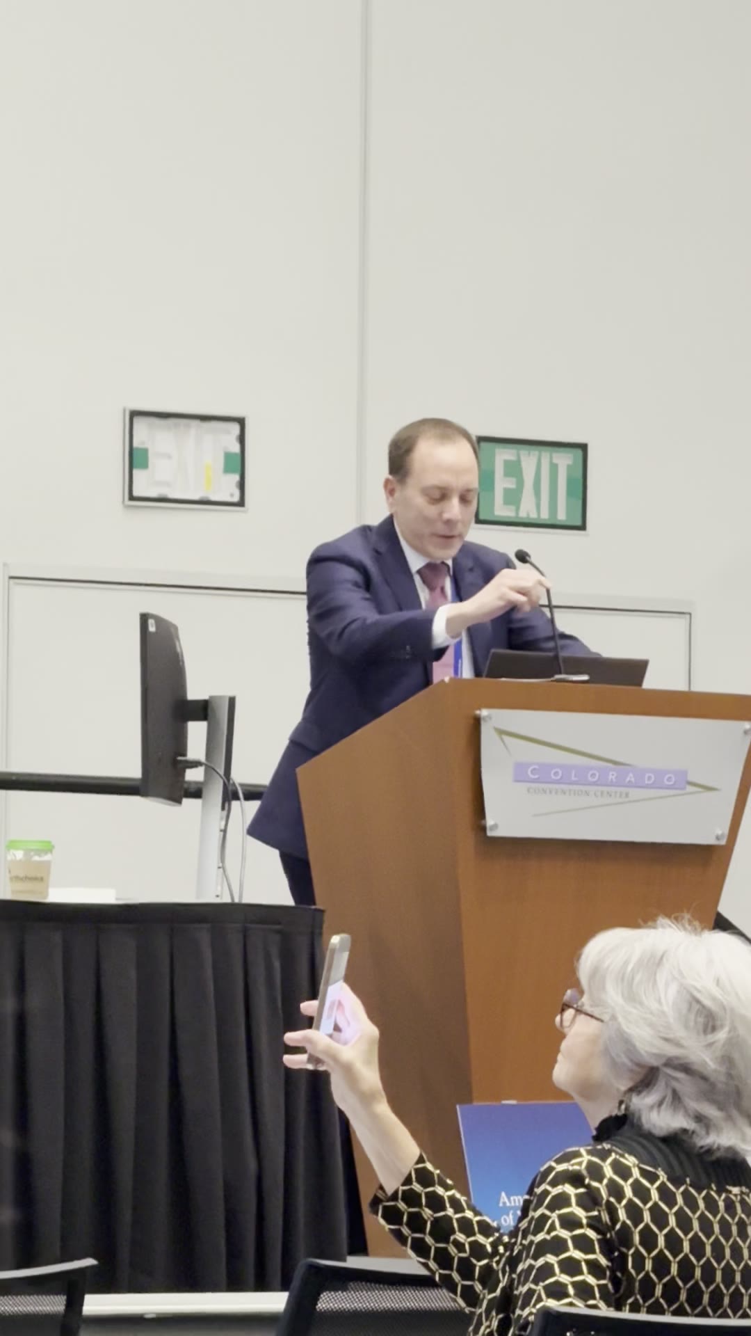
Ladd Medal Award Presentation Honoring Dr. Steven Rothenberg's Pioneering Minimally Invasive Pediatric Surgery
Dr. Steve Rothenberg · 33 min · Published May 2026
Video
PDA ligation by Dr. Steven Rothenberg
Dr. Steve Rothenberg · 6 min · Published Jun 2023
Video
Update Course Rewind: Pectus Excavatum 2021
Dr. Todd Ponsky · 11 min · Published Aug 2022
Podcast
Update Course Rewind: Magnet Therapy for Esophageal Atresia
10 min · Published Dec 2021
Video
Update Course Rewind: Thoracotomy vs VATS for Lung Metastases
Dr. Todd Ponsky · 9 min · Published Dec 2021
Video
Thoracoscopic TEF Repair, Is It Really Better Than Open? Update Course 2018
18 min · Published Aug 2018
Video
Intestinal Atresia Types Explained: Grossfeld Classification for Pediatric Surgery
1 min · Published Sep 2026
Video
Derivation and validation of the Pediatric Community-Acquired Pneumonia Severity (PedCAPS) score: A prospective cohort study
50 s · Published Sep 2026
Video
FETO for Late-Diagnosed Severe Congenital Diaphragmatic Hernia (CDH) at Cincinnati Children's with Dr. Beth Rymeski
4 min · Published Sep 2026
Video
Severe Congenital Diaphragmatic Hernia (CDH) Case: FETO Management & Unexpected Findings with Dr. Beth Rymeski
3 min · Published Sep 2026
Video
Fetoscopic Endoluminal Tracheal Occlusion (FETO) Procedure: Step-by-Step Guide with Dr. Beth Rymeski
4 min · Published Sep 2026
Video
Choledochal Cyst Types Explained: Pediatric Surgery Fundamentals
1 min · Published Sep 2026
What the experts said
The NEXUS trial criteria do not cover pediatric patients.
By NEXUS trial criteria, an 18-month-old with normal exam and no other risk factors should be clearable clinically, but the child's age makes clinical clearance unreliable.
C-spine injuries in very young children tend to be high cervical injuries, which may be missed on physical exam even when the child can move the neck and has no obvious tenderness.
MRI requires anesthesia in an 18-month-old, which is a barrier to obtaining it acutely.
One neck CT delivers radiation equivalent to 600 plain films.
The clinical significance of the radiation difference between plain films and CT is unknown; no one knows the true effects.
If a lateral C-spine film is inadequate, a CT will be needed, resulting in radiation exposure from both modalities.
In children 2 years and younger, a CT scan does not add diagnostic value above an adequate plain radiograph for C-spine evaluation.
In most institutions, an 18-month-old ejected from a vehicle would arrive as a level 2 trauma and automatically receive protocolized imaging including CT.
C-spine injuries in the 18-month age group almost always present with the child either dead or paralyzed.
A case was seen of an 18-month-old with spinal cord contusion and muscle highlighting on MRI that was missed on CT and had equivocal plain films; the child had no neurologic deficit but was not acting normal per mother.
SCIWORA (spinal cord injury without radiographic abnormality) can manifest hours after injury, with initial findings not present on early imaging.
Institutional C-spine clearance algorithms typically offer collar immobilization or MRI as reasonable final-step options, with the decision made by the clearing physician (orthopedics or neurosurgery).
Local protocol requires two-view (AP and lateral) C-spine films, not three-view; odontoid views are not obtained in young children.
There are two types of flexion-extension films: voluntary patient-directed movement in cooperative older children, and fluoroscopy-assisted movement performed by a physician (neurosurgery or trauma team) to avoid causing paralysis.
MRI is obtained in young children who cannot cooperate with voluntary flexion-extension films; older children receive either voluntary or assisted flexion-extension films.
Some patients with normal plain films and normal MRI have persistent midline tenderness; neurosurgery typically discharges these patients home in a soft collar for comfort, knowing the patient will remove it after about a week.
MRI is extremely accurate for ligamentous injury; when present, it is visible on MRI.
Over the last 5–10 years, work by colleagues has allowed age-stratified C-spine protocols; a 13-year-old is not treated like a 2-year-old.
Future work from PECARN will help determine which children do not need any C-spine imaging at all.
MRI is the gold standard for evaluating SCIWORA and ligamentous injury.
Plain films are the next step in a 10-year-old with midline tenderness, no distracting injuries, and GCS 15.
The degree of midline tenderness, believability of tenderness, and neck muscle spasm guide the astute clinician in deciding whether to proceed to MRI or observe in a collar.
Children without distracting injuries, without neurologic symptoms, and with minimal or no tenderness have been over-imaged; restraint in imaging this group is an area for improvement.
In children 8 years and older, high-risk features (midline tenderness, high mechanism) lead to skipping plain films and proceeding directly to CT, especially if a head CT is already indicated.
MRI is used for C-spine clearance in prolonged intubation or ICU patients despite a clear CT.
In a neurologically devastated patient, MRI is obtained at an elective time for C-spine clearance.
Even in an emergency requiring immediate surgery, if the head is scanned, the neck does not need to be scanned at that time; the patient will remain in a collar regardless of imaging results.
Extending a head CT through the neck takes only an extra 30 seconds and is faster than obtaining a separate plain radiograph.
Radiologists argue that the incremental radiation from extending a head CT through the neck is trivial, comparable to radiation from a transcontinental flight.