


Chapter 1 of 4 · Diagnosis & Workup
3D cloacography
3D Rotational Cloacography Technique and Technology
Expert statements on this page
No expert statements were drawn from this page.
Host summaries · secondary, not cited in answers
Educational content from recorded physician discussions — not medical advice. Talk to your (or your child's) care team about your situation.
Podcast
Colorectal Quiz Episode 17: Cloaca Part 1
27 min · Published Jul 2021
Podcast
Fetal and Newborn Management of Cloacal Malformations
13 min · Published Sep 2026
Video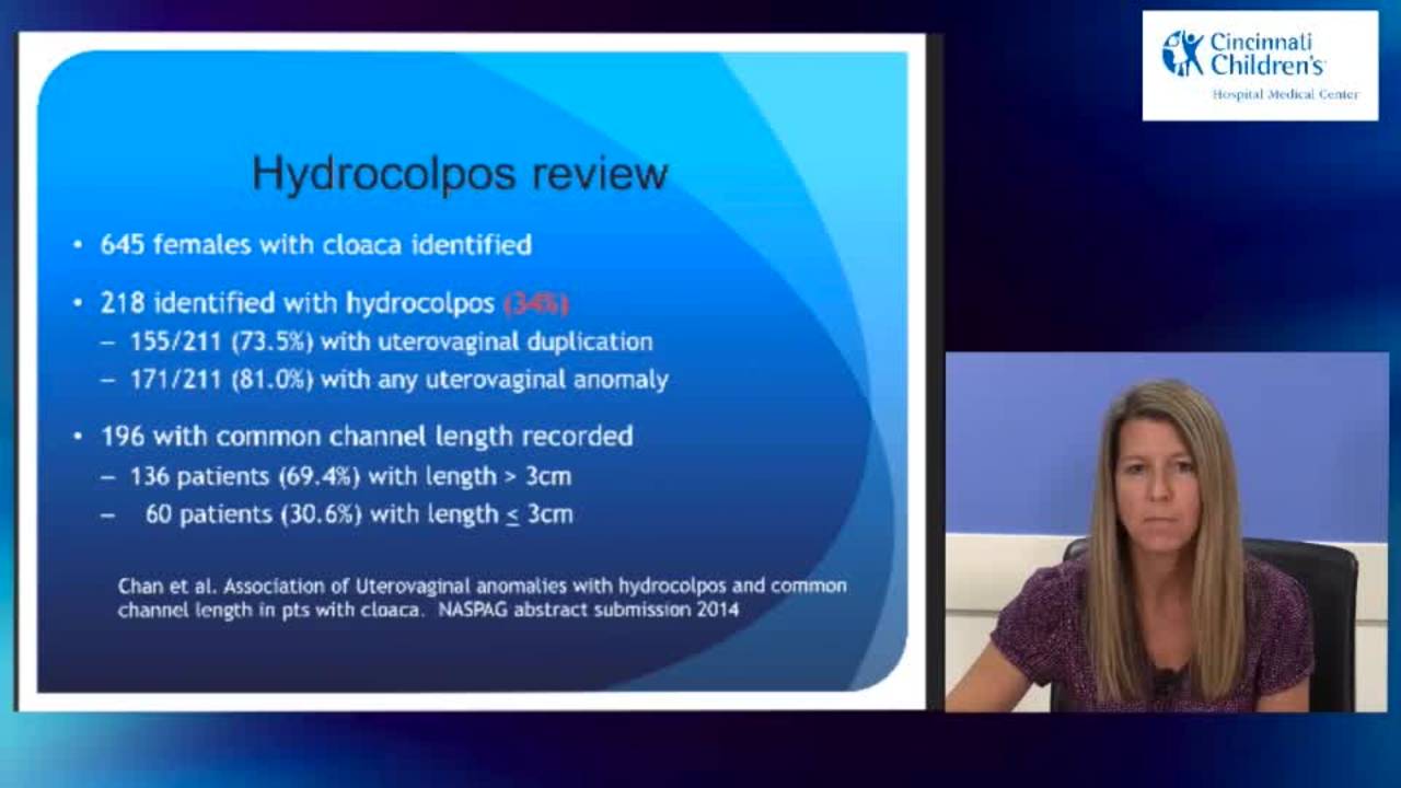
Case Presentations Part I: Cloaca and Complex ARMs 2015
45 min · Published Oct 2015
Podcast
Cloaca Management with Dr. Marc Levitt & Dr. Aaron Garrison
43 min · Published Dec 2016
Video
Cloaca - Case Presentations
45 min · Published Nov 2018
Video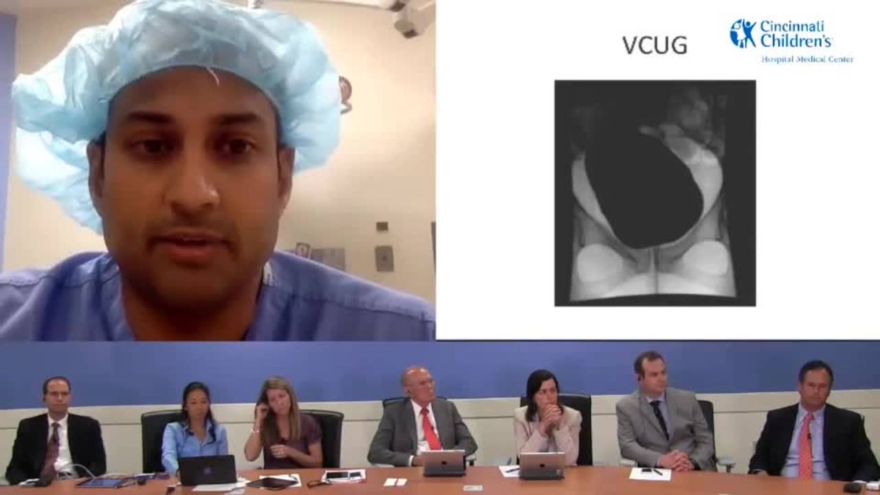
Surgical Management and Follow-Up: Cloaca and Complex ARMs 2015
25 min · Published Oct 2015
Video
Validation of an anorectal malformation trainer - Can a high-fidelity model simulate real life?
1 min · Published Jun 2026
Video
Complications and Long-Term Outcomes of Patients With Cloacal Malformation After Bowel Neovagina...
1 min · Published Jun 2026
Video
Safety and utility of long-acting steroid injection for management of post-operative stricture...
1 min · Published Jun 2026
Video
Association Between Social Determinants of Health and Choice of Urinary Reconstruction in Children
1 min · Published Jun 2026
Video
The Perineal Body Preserving PSARP (PPP)
11 min · Published Jun 2026
Video
Update Course Rewind 2025: Hirschsprung + ARM: Rare but Real
1 min · Published May 2026
Video
Intestinal Atresia Types Explained: Grossfeld Classification for Pediatric Surgery
1 min · Published Sep 2026
Video
Derivation and validation of the Pediatric Community-Acquired Pneumonia Severity (PedCAPS) score: A prospective cohort study
50 s · Published Sep 2026
Video
FETO for Late-Diagnosed Severe Congenital Diaphragmatic Hernia (CDH) at Cincinnati Children's with Dr. Beth Rymeski
4 min · Published Sep 2026
Video
Severe Congenital Diaphragmatic Hernia (CDH) Case: FETO Management & Unexpected Findings with Dr. Beth Rymeski
3 min · Published Sep 2026
Video
Fetoscopic Endoluminal Tracheal Occlusion (FETO) Procedure: Step-by-Step Guide with Dr. Beth Rymeski
4 min · Published Sep 2026
Video
Choledochal Cyst Types Explained: Pediatric Surgery Fundamentals
1 min · Published Sep 2026
What the experts said
3D rotational cloacography is performed in interventional radiology using angiography equipment, combining endoscopy with 3D imaging capabilities in a single session.
The 3D cloacography technique uses the same technology available for 3D angiography: contrast is injected into hollow structures while the camera rotates around the patient to generate images.
Images from 3D cloacography are immediately available at a workstation where different cuts and views can be made to clarify overlapping structures.
Conventional 2D cloacography with AP and lateral views has overlapping structures that make it difficult to decipher which structure is which.
Modern 3D cloacography technology allows precise measurement of the common channel, whereas the old method using an external ruler had significant foreshortening and imprecise measurements.
The 3D approach helps determine whether the vagina will come down and clarifies the relationship with the rectum, aiding decisions about laparoscopic versus open approaches.
The radiation dose from 3D rotational cloacography is very similar to conventional cloacography because fewer lateral and frontal images are needed.
MRI cloacography loses the real-time capability of seeing what is being injected and filled, and significantly increases anesthesia time.
The measurements from 3D cloacography are accurate to within millimeters; when the radiologist reports 3.5 cm, the intraoperative measurement is typically 3.5 cm.
The majority of cloacal malformations diagnosed prenatally are complex cases.
Baseline renal bladder ultrasound is the preferred method for identifying upper tract and lower tract urologic abnormalities in cloacal malformations.
Ultrasound is used to look for fluid accumulated in the vagina or vaginas, which helps guide the timing of therapy and whether fluid needs to be drained acutely.
Sacral X-ray (AP and lateral) is needed to assess prognosis for bowel control and to suspect or rule out a pre-sacral mass.
Spinal ultrasound is performed to rule out tethered cord in cloacal malformation workup.
Placement of a catheter into the common channel is challenging because it may go into any of three organ systems (bladder, vagina, or rectum), making standard VCUG unreliable.
High-grade reflux is not commonly identified in cloacal malformation patients; most reflux is mild and warrants observation rather than mandatory preoperative intervention.
In neonatal cloacas, trying to make a very accurate anatomic diagnosis during the newborn period is useless and may actually hurt the baby.
In the neonatal period, the crucial information is whether the baby has hydronephrosis, megaureters, or hydrocolpos compressing the ureters, which can be determined by ultrasound without endoscopy.
Knowing whether a neonate with cloaca has reflux is not crucial; the priority is decompressing the gastrointestinal and urogenital tracts.
Forcing an endoscope into a tiny neonatal cloacal structure can cause harm.
Nephrostomy, ureterostomy, and vesicostomy are not indicated in most neonatal cloacas.
The recommended neonatal management for cloaca is to drain hydrocolpos, open a colostomy, and re-evaluate from the urologic point of view 48 hours later before making further decisions.
Some cloacal patients may need vesicostomy if there is obstruction in the common channel, but many do not; decompressing the hydrocolpos often completely changes the clinical picture.
Neonatal endoscopy of cloacas does not always provide accurate anatomic information and can be traumatizing to the common channel, potentially causing harm.