


From
Live Event Content
2024 Fetal Care Center Frontiers in Fetal Neurology Day 1 - Dr. Dawn Gano
Chapter 1 of 6 · Fundamentals
Hydrocephalus pathophysiology
Introduction and pathophysiology of obstructive hydrocephalus
Expert statements on this page
No expert statements were drawn from this page.
Host summaries · secondary, not cited in answers
Educational content from recorded physician discussions — not medical advice. Talk to your (or your child's) care team about your situation.
Video
2024 Fetal Care Center Frontiers in Fetal Neurology Day 1 - Dr. Jose Peiro
16 min · Published Oct 2024
Video
2024 Fetal Care Center Frontiers in Fetal Neurology Day 1 - Hour 1 Q&A
9 min · Published Oct 2024
Video
2024 Fetal Care Center Frontiers in Fetal Neurology Day 1 - Dr. Jesse Skoch
19 min · Published Oct 2024
Podcast
IFMSS 2019 Techincal & Educational Innovation
14 min · Published Nov 2022
Video
Embryology Essentials For FRCS Neurosurgery Exam
Amr Moursi · 15 min · Published Dec 2024
Video
Update Course Rewind: Highlighting Collaboration from Surgery & Interventional Radiology in the OR 2024
CCHMC Pediatric Surgery · 10 min · Published Mar 2025
Video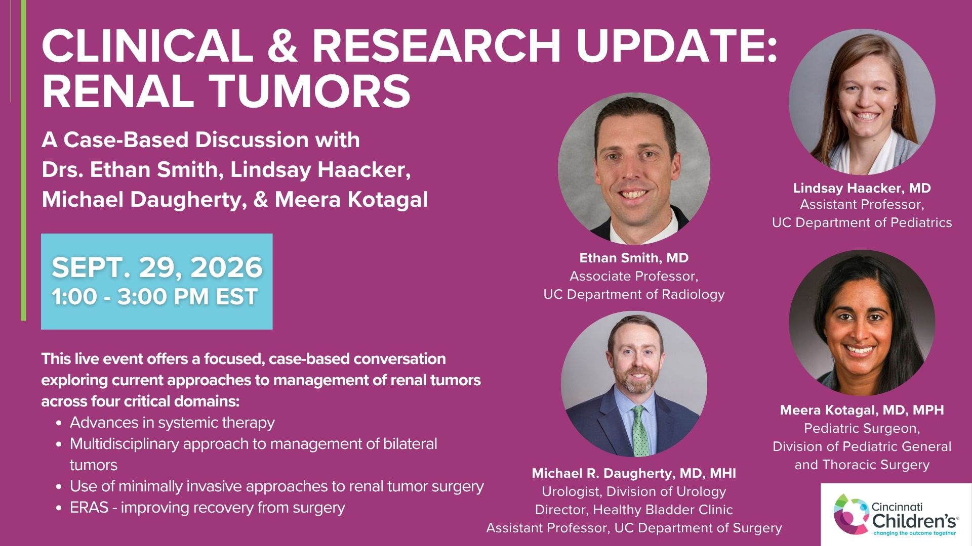
Clinical & Research Update: Renal Tumors with Drs. Ethan Smith, Lindsay Haacker, Michael Daugherty, and Meera Kotagal
71 min · Published Sep 2026
Video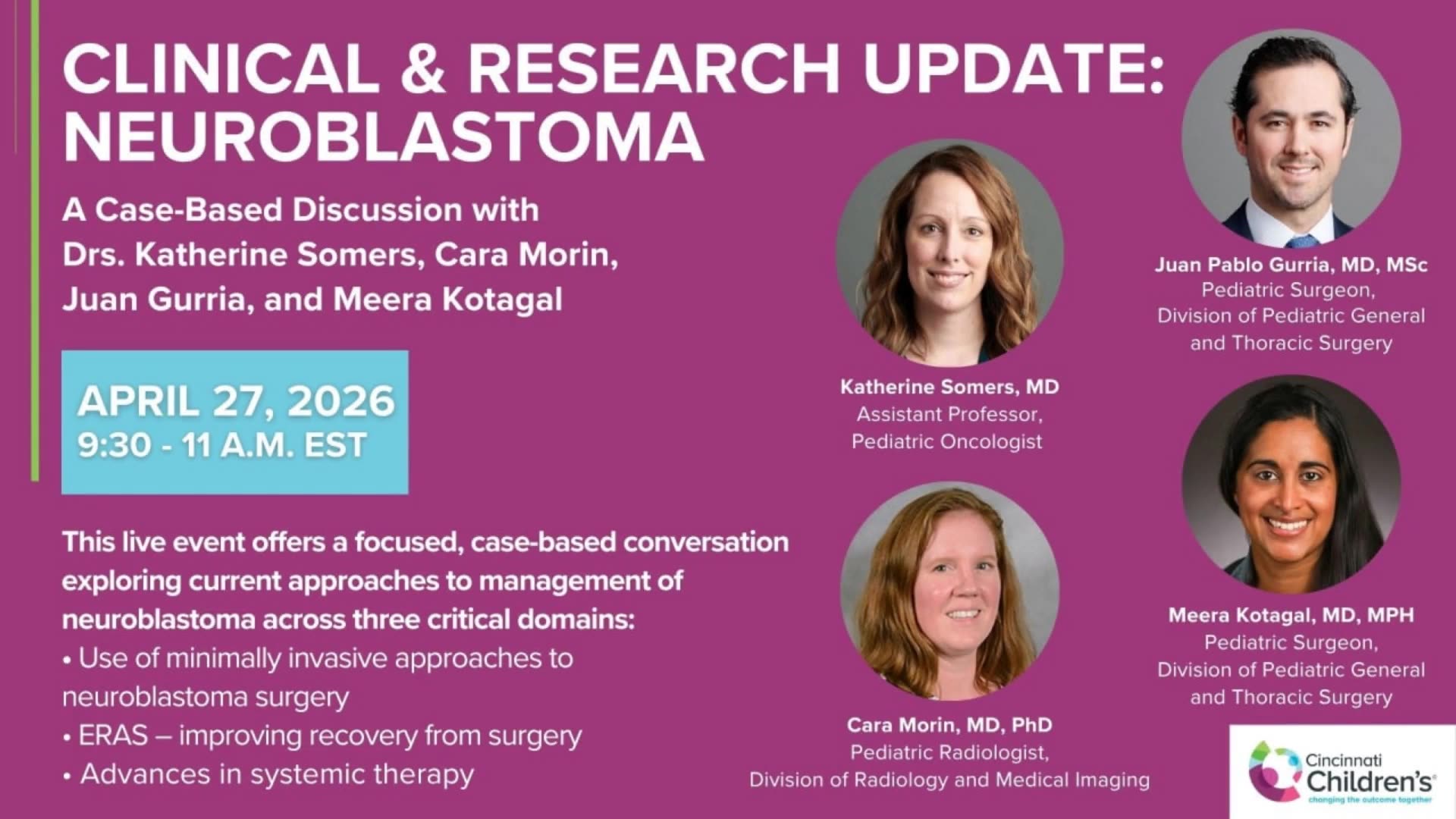
Clinical & Research Update: Neuroblastoma with Drs. Katherine Somers, Cara Morin, Juan Gurria, and Meera Kotagal
67 min · Published Sep 2026
Video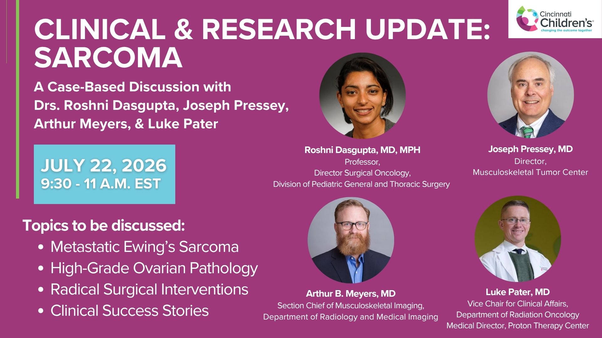
Clinical & Research Update: Sarcoma w/ Drs. Roshni Dasgupta, Joseph Pressey, Arthur Meyer, Luke Pater
66 min · Published Sep 2026
Video
2026 Laparoscopic Pediatric Hernia Repair
123 min · Published Jun 2026
Video
Beyond the Spectrum: Diagnosis, Myths & Management - Caitlin Couch & Leslie Lopez - APP Conference 2026
50 min · Published May 2026
Video
Dysautonomia: Navigating the Journey - Martha Willis - APP Conference 2026
55 min · Published May 2026
What the experts said
CSF is produced in the choroid plexus and circulates through the ventricular system, through the aqueduct of Sylvius and fourth ventricle, around the brain for reabsorption.
Aqueductal stenosis blocks normal CSF circulation and causes fluid buildup in the lateral and third ventricles, representing a pure obstructive problem in fetuses and babies.
Progressive ventricular enlargement in obstructive hydrocephalus leads to increased intracranial pressure, cortical mantle thinning, rupture of the septum pellucidum, and denudation of the ependyma, causing problems in brain development.
Ventriculomegaly and hydrocephalus can be detected very early in gestation, with severity classified as mild, moderate, or severe.
MRI is a very good tool to complete prenatal diagnosis and identify isolated aqueductal stenosis, along with genetic testing.
Aqueductal stenosis is the most important cause of obstructive congenital hydrocephalus.
Babies with congenital hydrocephalus almost consistently need CSF diversion, mostly with VP shunts, which have problems including malfunction and infection.
Postnatal treatment of hydrocephalus probably arrives late because progressive brain deterioration occurs during the intrauterine period, including denudation, astrocytosis, and microgliosis.
The first attempts at fetal hydrocephalus treatment were in fetal monkeys using ventriculo-amniotic shunts approximately 40 years ago.
The first human case of ventriculo-amniotic shunt was described by Birnholz almost 40 years ago.
A registry collected 44 cases of ventriculo-amniotic shunts, and results discussed at a 1982 fetal medicine and surgery meeting led experts to declare a moratorium on this intervention due to inadequate clinical outcomes and patient selection.
After 40 years, improved technology, imaging, and genetic tools for patient selection make it reasonable to reconsider fetal hydrocephalus treatment, but using ETV rather than shunts.
In the fetal lamb model, kaolin injected into cisterna magna creates hydrocephalus but produces a meningitis-like irritation reaction.
Comparing kaolin, onyx, and bio-glue as agents to create hydrocephalus in fetal lambs, bio-glue was determined to be the best.
Bio-glue was the only agent that allowed creation of severe hydrocephalus cases in the fetal lamb model.
Brain thinning was more significant with bio-glue compared to other agents in the fetal lamb hydrocephalus model.
The fetal lamb hydrocephalus model shows ependymal denudation with loss of cilia, likely due to ventricular distension.
The percentage of ependymal denudation is higher with more severe hydrocephalus in the fetal lamb model.
Ependymal denudation occurs very early in gestation and persists until delivery in the fetal lamb model.
Astrocytes replace ependymal cells and create scar tissue in areas of denudation in the hydrocephalus model.
Activated microglia indicating neuroinflammation is present in areas of ependymal denudation, putting neuroprogenitor cells in the subventricular zone at risk of maldevelopment.
A 7 French rigid fetoscope or cystoscope was used to perform fetal ETV in the lamb model after trying different scopes including a yellow scope.
The entry point for fetal ETV in lambs is at the coronal suture on the right side, approximately 1 centimeter to the midline to enter the ventricle.
Fetal ETV can be performed transuterine without opening the uterus in the lamb model.
The fetal lamb brain anatomy is more elongated than human, with a narrower foramen of Monro, creating technical difficulties for fetal ETV.
In the fetal lamb model, there is no septum pellucidum, allowing visualization of both sides of the lateral ventricles.
The interthalamic adhesion is very large in fetal lambs, making the anatomy different from humans.
A rigid plastic fiber (a laser without energy) is used to create a blunt opening in the third ventricle floor to avoid damaging the basilar artery in mid-gestation fetuses.
In twin fetal lambs with the same degree of ventriculomegaly, the one treated with fetal ETV had thicker brain tissue and smaller ventriculomegaly at delivery compared to the untreated twin.
A fetal surgeon in Brazil has begun performing fetal ETV in humans percutaneously with a sharp-tip cystoscope and has published 10 cases.
Fetal ETV in the lamb model can reduce ventricular size from severe to moderate-mild and from moderate to normal.
Fetal ETV can improve cortical thickness in the fetal lamb hydrocephalus model.
Fetal ETV is feasible in both fetal lambs and humans.
In most cases in the lamb model, fetal ETV recovered ependymal denudation, though it is unknown whether this represents prevention or regeneration after ventricular deflation.
Fetal ETV is safe with no deaths reported in the Brazilian series of 10 human cases.
Plans exist for a multi-center US clinical trial with phase one enrolling 10 cases to assess safety and feasibility, followed by phase two enrolling 50-60 cases to assess efficacy.
Fetal hydrocephalus is associated with potentially poor outcomes including intellectual disabilities, spasticity, and seizures.
Endoscopic third ventriculostomy can be performed postnatally to recirculate CSF as an alternative to shunting.