


From
Live Event Content
2024 Fetal Care Center Frontiers in Fetal Neurology Day 1 - Dr. Charu Venkatesen
With Dr. Charu Venkatesen
Chapter 1 of 5 · Fundamentals
CAS introduction
Introduction to Congenital Aqueductal Stenosis and Ventriculomegaly
Expert statements on this page
No expert statements were drawn from this page.
Host summaries · secondary, not cited in answers
Educational content from recorded physician discussions — not medical advice. Talk to your (or your child's) care team about your situation.
Video
2024 Fetal Care Center Frontiers in Fetal Neurology Day 1 - Introduction
2 min · Published Oct 2024
Video
2024 Fetal Care Center Frontiers in Fetal Neurology Day 1 - Dr. Jesse Skoch
19 min · Published Oct 2024
Video
2024 Fetal Care Center Frontiers in Fetal Neurology Day 1 - Hour 1 Q&A
9 min · Published Oct 2024
Only a few other public items share this expert — go deeper there →
Video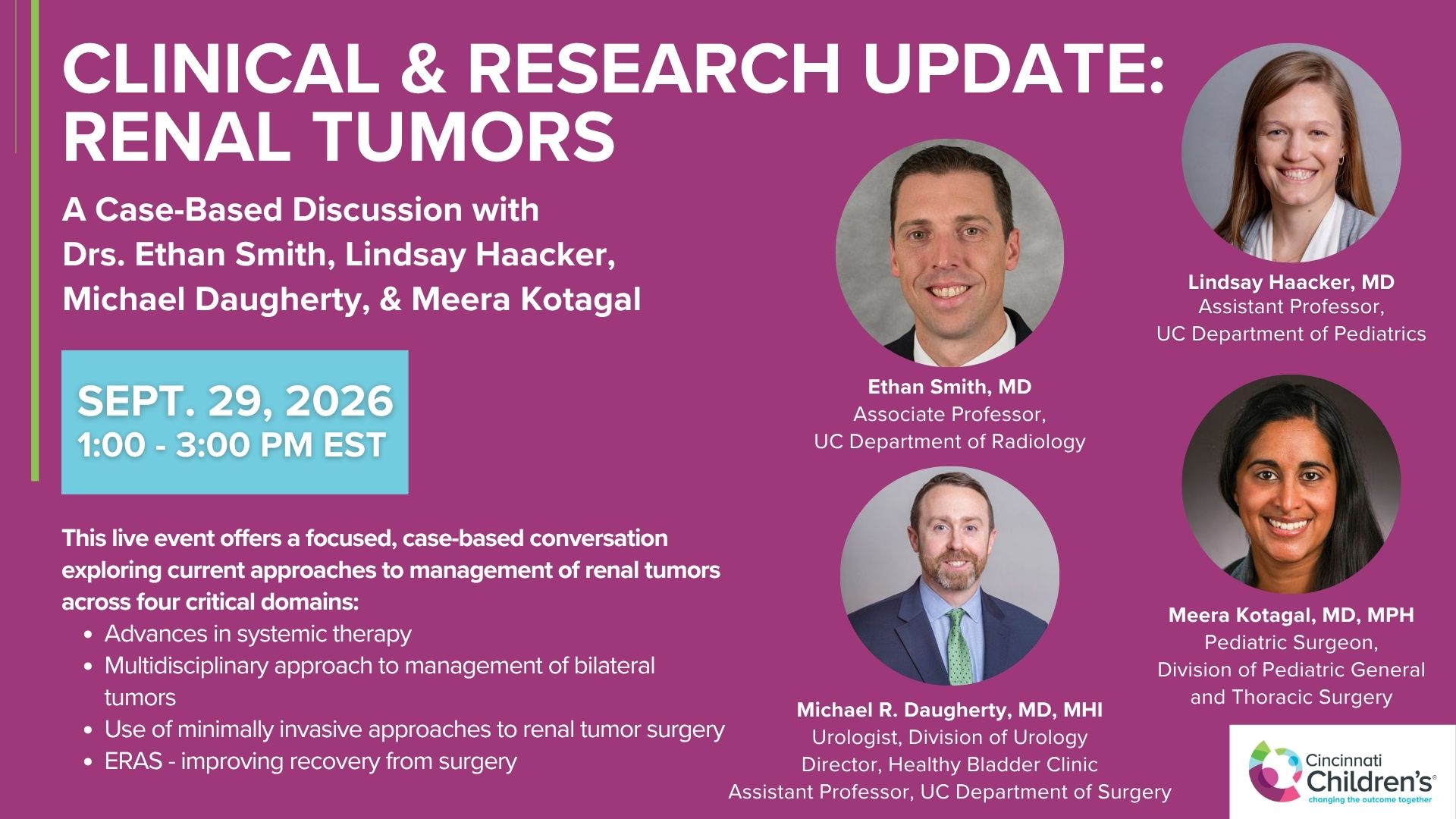
Clinical & Research Update: Renal Tumors with Drs. Ethan Smith, Lindsay Haacker, Michael Daugherty, and Meera Kotagal
71 min · Published Sep 2026
Video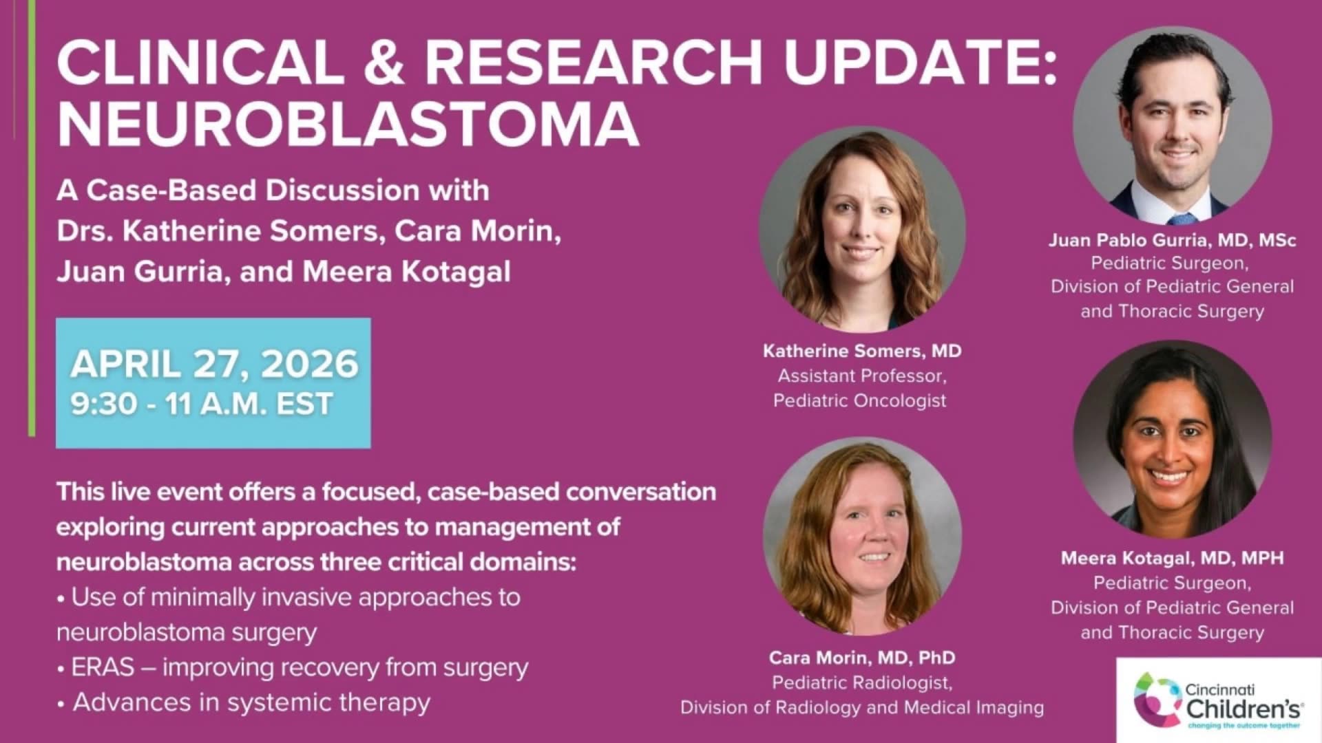
Clinical & Research Update: Neuroblastoma with Drs. Katherine Somers, Cara Morin, Juan Gurria, and Meera Kotagal
67 min · Published Sep 2026
Video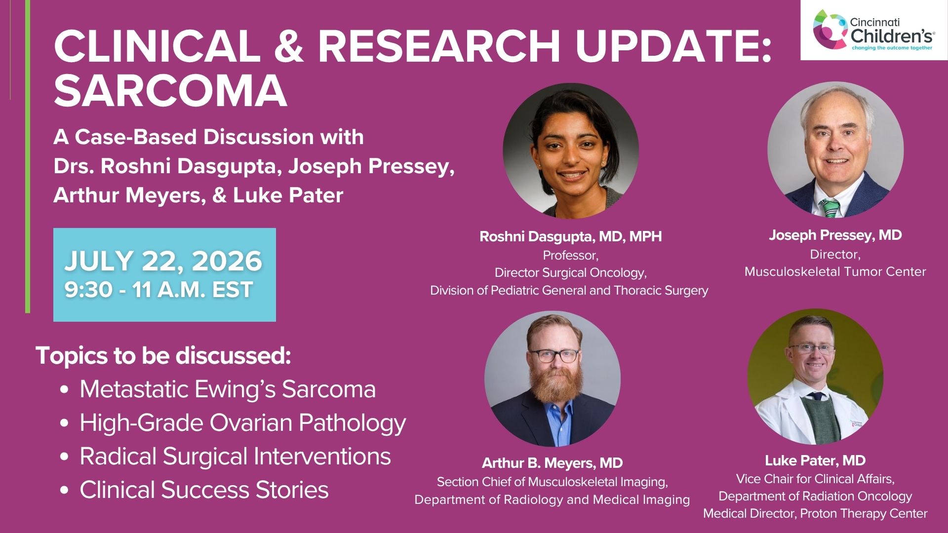
Clinical & Research Update: Sarcoma w/ Drs. Roshni Dasgupta, Joseph Pressey, Arthur Meyer, Luke Pater
66 min · Published Sep 2026
Video
2026 Laparoscopic Pediatric Hernia Repair
123 min · Published Jun 2026
Video
Beyond the Spectrum: Diagnosis, Myths & Management - Caitlin Couch & Leslie Lopez - APP Conference 2026
50 min · Published May 2026
Video
Dysautonomia: Navigating the Journey - Martha Willis - APP Conference 2026
55 min · Published May 2026
What the experts said
Ventriculomegaly is the most common CNS abnormality identified on fetal imaging.
Fetal ventriculomegaly is measured at the atrium of the lateral ventricles, with anything larger than 10 millimeters considered abnormal: 10-12mm is mild, 12.1-15mm is moderate, and over 15mm is severe.
By the time patients with congenital aqueductal stenosis come to a fetal care center, their ventricular measurements are typically in the moderate or severe range.
Congenital aqueductal stenosis on MRI is defined by a thin corpus callosum, thickened tectum, fluid through the aqueduct not seen all the way through with narrowing and obstruction at the inferior portion of the aqueduct, and a large third ventricular recess.
The incidence of congenital aqueductal stenosis ranges from 0.5 to 1 per 1000 births.
Congenital aqueductal stenosis etiology can be acquired (intrinsic obstruction from post-hemorrhagic insult or infection causing gliosis, or extrinsic compression from a mass) or due to genetic disorders including L1CAM mutation, rhombencephalosynapsis, and distroglycanopathies such as Walker-Warburg syndrome.
In the Cincinnati study of 140 cases of congenital aqueductal stenosis identified by fetal MRI, 16% had early demise (termination or fetal demise), 3% were lost to follow-up, and 113 were live births.
Of 113 live births in the Cincinnati CAS study, 11% died prior to hospital discharge and 8% were lost to follow-up, leaving short-term neurodevelopmental data available for 86 patients.
Isolated congenital aqueductal stenosis is defined as CAS being the only anomaly on imaging with no other intra or extracranial findings, while complex CAS includes any accompanying intra or extracranial anomalies or genetic abnormalities.
At time of discharge in the Cincinnati study, 95% of complex CAS cases required CSF diversion (all via ventriculoperitoneal shunt) versus 70% of isolated CAS cases.
At discharge, 63% of complex CAS cases required feeding support versus 29% of isolated CAS cases.
There were no differences between isolated and complex CAS groups in terms of seizures or respiratory support at discharge.
For long-term motor outcomes, 93% of isolated CAS patients were fully ambulatory versus 44% of complex CAS patients.
None of the isolated CAS patients were non-ambulatory long-term, whereas 32% of complex CAS patients were non-ambulatory.
Long-term epilepsy rates were 56% in complex CAS versus 20% in isolated CAS.
Long-term feeding support via gastrostomy tube was required in approximately five times as many complex CAS cases compared to isolated CAS cases.
If you have isolated CAS, you have very favorable motor outcome.
International registries are needed for congenital aqueductal stenosis and other rare prenatally identified neurological conditions to accumulate hundreds to thousands of patients.
Many prenatally identified neurological conditions including agenesis of the corpus callosum and vermian hypoplasia can be categorized as isolated or complex, but complex is a very heterogeneous category requiring better genotype-phenotype characterization.
Longitudinal studies are needed to characterize the adult-lived experience beyond age 10, including patient and family-defined outcomes such as stressors on families, perception of pain, quality of life, and independence.
More studies are needed examining postnatal and fetal interventions to improve outcome in congenital aqueductal stenosis.
In the literature, postnatal mortality for congenital aqueductal stenosis is reported to range from 34 to 68%.
In the literature, typical development in congenital aqueductal stenosis is reported to range from 4% to 63%.
Epilepsy risk in congenital aqueductal stenosis is reported around 50% in the literature.