


From
StayCurrentMD
Laparoscopic Intestinal Duplication Cyst Repair - Technique
With Dr. Steve Rothenberg
Chapter 1 of 4 · Case-Based Learning
Case & setup
Case presentation and initial exploration
Expert statements on this page
No expert statements were drawn from this page.
Host summaries · secondary, not cited in answers
Educational content from recorded physician discussions — not medical advice. Talk to your (or your child's) care team about your situation.
Video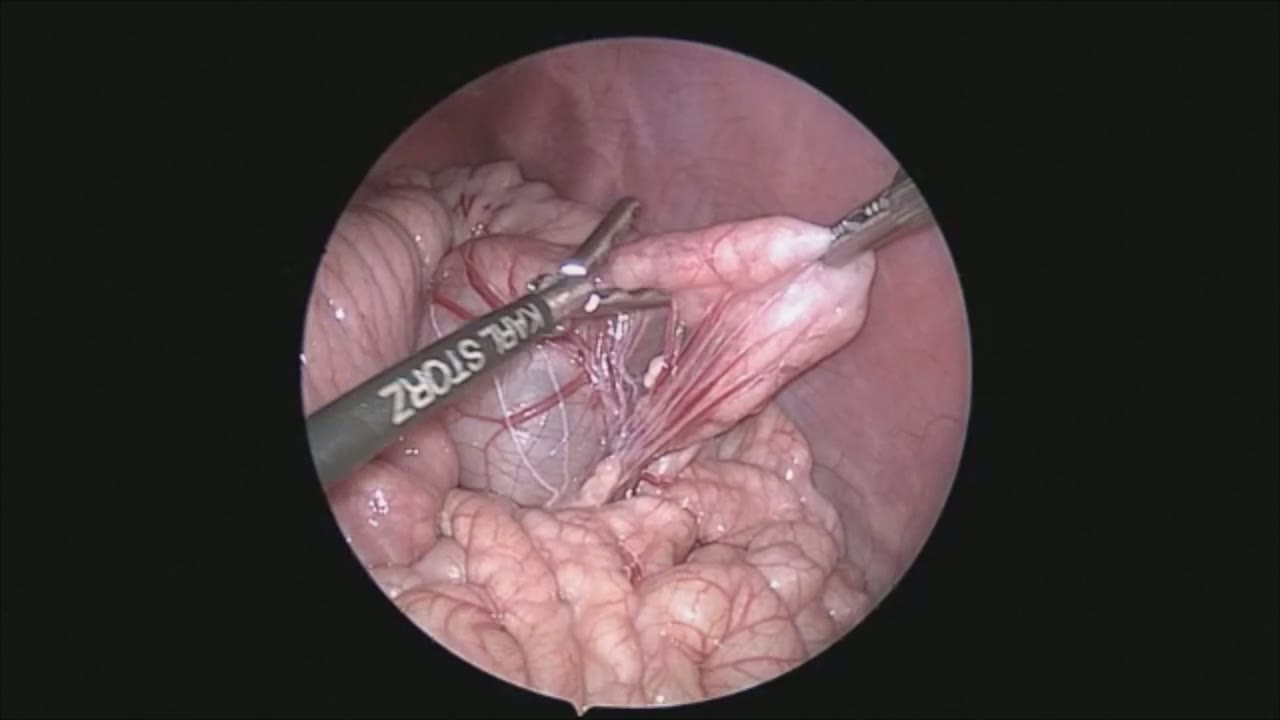
sealer demo
Dr. Steve Rothenberg · 3 min · Published May 2026
Video
pyloric knife
Dr. Steve Rothenberg · Published May 2026
Video
pulm artery LLL
Dr. Steve Rothenberg · 9 s · Published May 2026
Video
plication narrated
Dr. Steve Rothenberg · 6 min · Published May 2026
Video
percuvance redo nissen
Dr. Steve Rothenberg · 5 min · Published May 2026
Video
open bowel anast
Dr. Steve Rothenberg · Published May 2026
Video
Intestinal Atresia Types Explained: Grossfeld Classification for Pediatric Surgery
1 min · Published Sep 2026
Video
Derivation and validation of the Pediatric Community-Acquired Pneumonia Severity (PedCAPS) score: A prospective cohort study
50 s · Published Sep 2026
Video
FETO for Late-Diagnosed Severe Congenital Diaphragmatic Hernia (CDH) at Cincinnati Children's with Dr. Beth Rymeski
4 min · Published Sep 2026
Video
Severe Congenital Diaphragmatic Hernia (CDH) Case: FETO Management & Unexpected Findings with Dr. Beth Rymeski
3 min · Published Sep 2026
Video
Fetoscopic Endoluminal Tracheal Occlusion (FETO) Procedure: Step-by-Step Guide with Dr. Beth Rymeski
4 min · Published Sep 2026
Video
Choledochal Cyst Types Explained: Pediatric Surgery Fundamentals
1 min · Published Sep 2026
What the experts said
A full-term infant with a prenatally diagnosed abdominal cyst underwent ultrasound which confirmed the presence of a duplication cyst.
At 3 weeks of age, the patient underwent laparoscopic exploration.
The surgeon is positioned at the patient's feet for this procedure.
3 ports were used with ports in the right and left mid quadrant used for the manipulation and resection.
The initial maneuver was to elevate the transverse colon in order to run the bowel.
The intestinal duplication cyst appeared to be mid-jejunal.
The left mid quadrant port was upsized to a 5 millimeter port to allow for placement of the endoscopic stapler.
A single load of the stapler was used to divide the bowel both on the proximal and then on the distal end.
This allowed for division of the bowel without any intraabdominal contamination from the bowel contents.
The 3 millimeter vessel sealer was used to seal the mesentery and then it was stripped off the cyst in the bowel without any bleeding or other issues.
For vessels of this size, this maneuver is extremely efficient as it allows for rapid sealing and division of the mesentery without the repeated placement of endoscopic scissors.
A 3-0 Prolene stitch was placed through the anterior abdominal wall and then placed through first the distal limb and then the proximal limb of the bowel to align the two segments for the side to side anastomosis.
A Vicryl suture is placed intracorporeally to align the proximal segments of the two pieces of bowel to facilitate the side to side anastomosis.
Two enterotomies were then made side by side in each limb of the bowel to allow for placement of the 5 millimeter stapler.
A side to side anastomosis was completed with a single application of the stapler.
The resultant enterotomy was then closed with a running 4-0 Vicryl suture.
This was a very fast and efficient way of closing the small enterotomy.
The surgeon can follow himself, keeping adequate tension on the running suture line to prevent any gaps.
A small specimen bag was placed into the abdomen, which was the thumb of a #8 glove.
The cyst was decompressed using cautery and suction to allow the bowel to fit in the specimen bag.
The specimen was brought out through the left mid quadrant trocar site.