


From
Live Event Content
BOB Ped Surg 2023 - Rustam Yuldashev, WOFAPS - Presentation
With Dr. Rustam Yuldashev
Chapter 1 of 5 · Fundamentals
Portal cavernoma background
Introduction and Background on Portal Cavernoma
Expert statements on this page
No expert statements were drawn from this page.
Host summaries · secondary, not cited in answers
Educational content from recorded physician discussions — not medical advice. Talk to your (or your child's) care team about your situation.
Video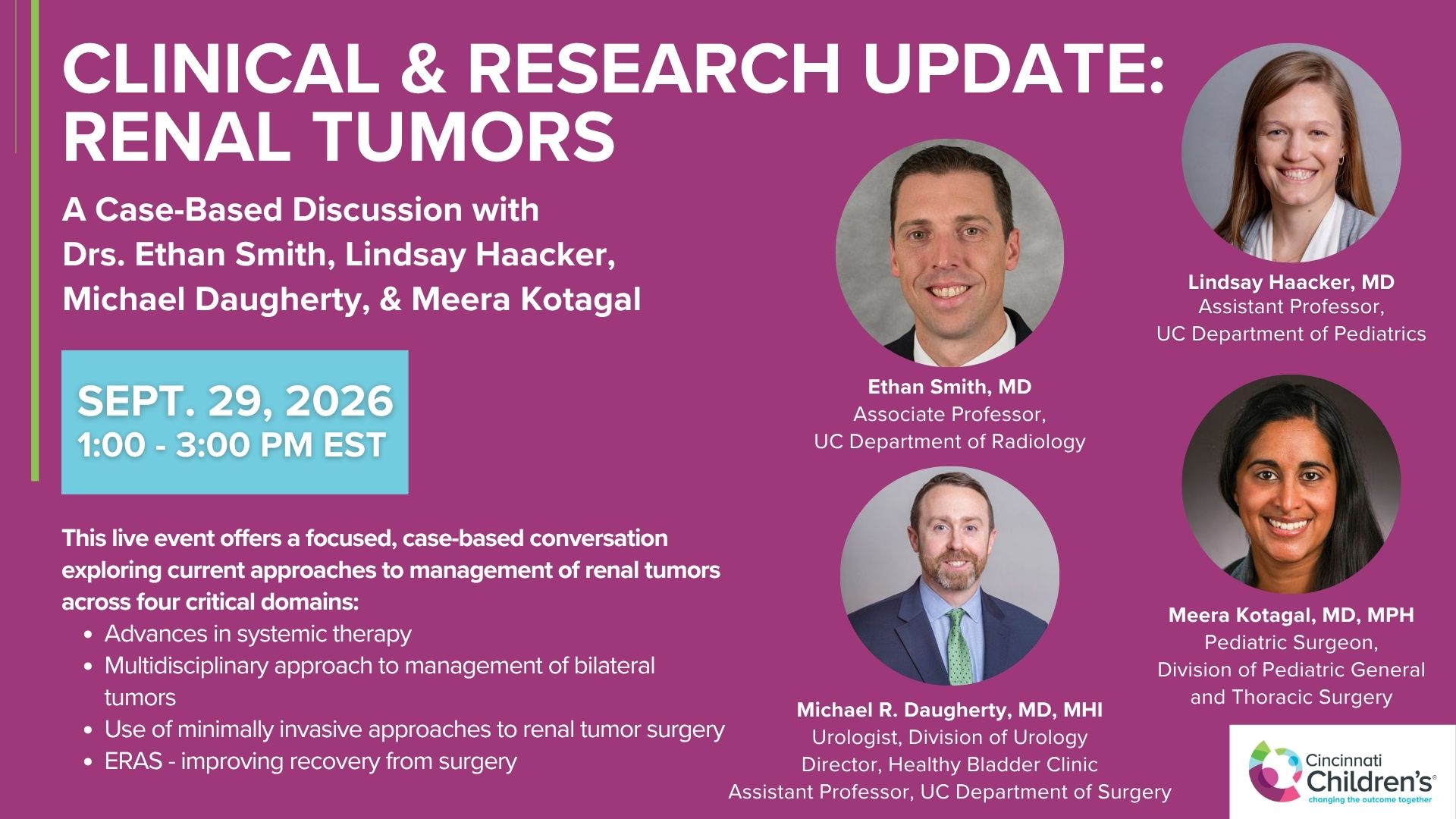
Clinical & Research Update: Renal Tumors with Drs. Ethan Smith, Lindsay Haacker, Michael Daugherty, and Meera Kotagal
71 min · Published Sep 2026
Video
Clinical & Research Update: Neuroblastoma with Drs. Katherine Somers, Cara Morin, Juan Gurria, and Meera Kotagal
67 min · Published Sep 2026
Video
Clinical & Research Update: Sarcoma w/ Drs. Roshni Dasgupta, Joseph Pressey, Arthur Meyer, Luke Pater
66 min · Published Sep 2026
Video
2026 Laparoscopic Pediatric Hernia Repair
123 min · Published Jun 2026
Video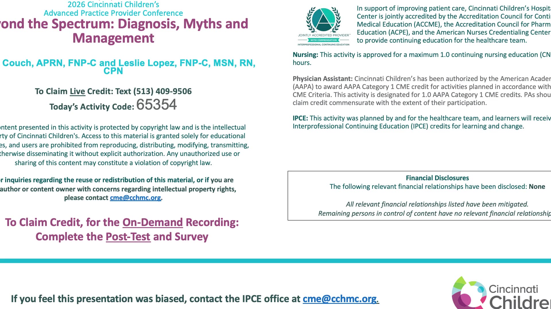
Beyond the Spectrum: Diagnosis, Myths & Management - Caitlin Couch & Leslie Lopez - APP Conference 2026
50 min · Published May 2026
Video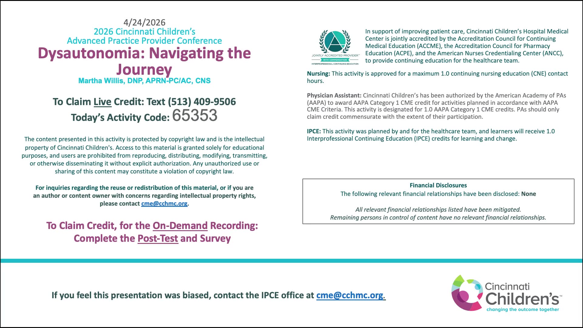
Dysautonomia: Navigating the Journey - Martha Willis - APP Conference 2026
55 min · Published May 2026
What the experts said
Majority of children with extrahepatic portal hypertension have isolated portal vein obstruction replaced by multiple venous collaterals (portal cavernoma).
Children with portal cavernoma often present with splenomegaly, thrombocytopenia, esophageal and gastric varices, and GI bleeding.
Balfour and Stewart first described cavernous transformation of the portal vein in 1869.
In the majority of patients with portal cavernoma, the etiology remains unknown even today.
155 children with portal vein obstruction were included in the study with a median age at evaluation of 9 years.
The majority of children with extrahepatic portal vein obstruction had pathological conditions in the neonatal period: omphalitis, musculoskeletal septic conditions, prolonged jaundice, and prematurity were noted with almost equal frequency.
Among known etiological factors, umbilical vein catheterization was most frequent.
In 48% of cases, children with portal vein obstruction had no identifiable etiological factors.
Five imaging patterns of portal-mesenteric obstruction were defined: type 1 (occlusion at portal vein bifurcation with patent SMV and splenic vein), type 2 (occlusion at main portal vein trunk with patent SMV and splenic vein), type 3 (portal vein occlusion extended to splenic vein), type 4 (portal vein and SMV occlusion with patent splenic vein), and type 5 (widespread thrombosis of the portal venous system).
The most frequent patterns were type 1 and type 5; the least common was type 3.
Type 5 pattern (widespread thrombosis) was the worst from a surgical point of view.
Children with type 1 and type 2 patterns had higher incidence of umbilical vein catheterization in the neonatal period.
Children with type 5 pattern (widespread thrombosis) had higher incidence of musculoskeletal septic conditions in the neonatal period.
Children with type 1 and type 3 patterns were admitted to hospital significantly earlier than children in other groups.
The correlation between age at presentation and type of portal vein obstruction was weak.
Children with type 3 and type 5 patterns had the highest incidence of GI bleeding episodes.
Children with type 3 and type 5 patterns had higher incidence of gastric varices.
Children with type 4 pattern (mesenteric vein thrombosis) never had portal hypertensive gastropathy.
Children with type 4 pattern had lower incidence of gastric varices and lower rate of cherry red spots on endoscopy.
Children with type 4 pattern had significantly lower grades of esophageal varices, especially compared to type 1 and type 2 patterns.
Thrombocytopenia was a common clinical feature of type 3 pattern and was observed in all cases.
Significant differences in platelet concentration were found only between children with type 4 and type 5 patterns.
The most common angiographic pattern of extrahepatic portal vein obstruction in children is type 1 (obstruction at the level of the bifurcation of the portal vein).
Children with splenic vein thrombosis (type 3) and widespread thrombosis (type 5) had the most severe clinical manifestations.
Children with mesenteric vein thrombosis (type 4 pattern) are characterized by lower risk of bleeding from esophageal varices and less incidence of gastric varices on GI endoscopy.