


Chapter 1 of 5 · Fundamentals
Pulmonary hypertension
Giant omphalocele physiology and pulmonary hypertension risk
Expert statements on this page
No expert statements were drawn from this page.
Host summaries · secondary, not cited in answers
Educational content from recorded physician discussions — not medical advice. Talk to your (or your child's) care team about your situation.
Podcast
Abdominal Wall Defects with Dr. Jacob Langer
52 min · Published Dec 2016
Video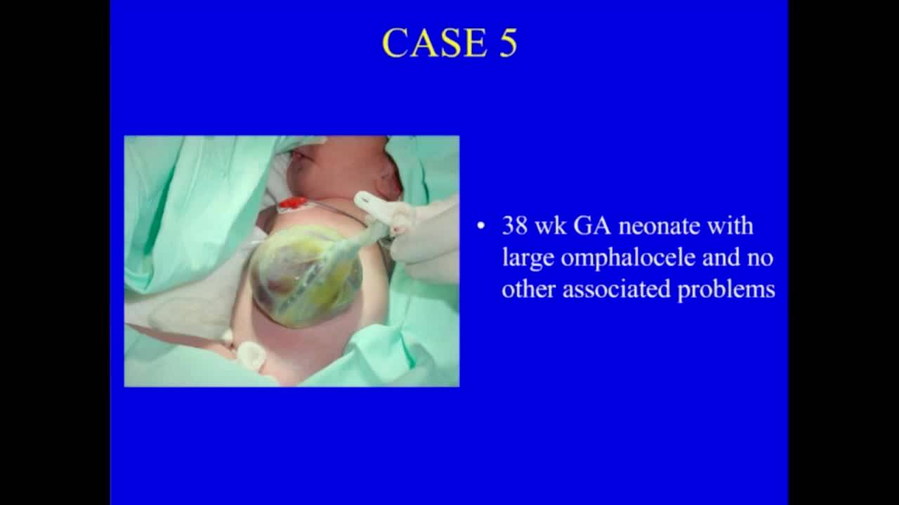
Abdominal Wall Defects
37 min · Published Nov 2018
Video
Abdominal Wall Defects: Update Course 2013
37 min · Published Sep 2013
Podcast
Malrotation with Dr. Meera Kotagal
13 min · Published May 2023
Video
Antibiotic Stewardship - APSA Practice Gaps 2019
Published Mar 2020
Video
Omphalocele & Gastroschisis
Dr. Todd Ponsky · 9 min · Published Mar 2022
Video
Impact of social determinants of health on outcomes in pediatric short bowel syndrome...
43 s · Published Aug 2026
Video
Advancements in Pediatric Intestinal Failure: Innovative Therapies and Improved Outcomes
17 min · Published Aug 2026
Video
Update Course Rewind 2025: Updates in NEC Management
11 min · Published Jul 2026
Video
Availability, utilization, and barriers to bowel ultrasound for necrotizing enterocolitis...
55 s · Published Jul 2026
Video
Malrotation and Volvulus with Trinity
Dr. Steve Rothenberg · 8 min · Published May 2026
Video
Malrotation Infant
Dr. Steve Rothenberg · 9 min · Published May 2026
Podcast
Update Course Rewind: Management of Esophageal Leaks 2022
Miguel Guelfand · 8 min · Published Jul 2026
Podcast
Update Course Rewind: Omphalocele & Gastroschisis 2020
Miguel Guelfand · 15 min · Published Jul 2026
Video
Pediatric Surgeon Miguel Guelfand, MD
Miguel Guelfand · Published Jul 2026
Video
Atresia de Esófago Compleja Dr. Miguel Guelfand
Miguel Guelfand · Published Jul 2026
Video
Atresia Esofágica sin Fístula Long Gap - Dr. Miguel Guelfand Clínica Las Condes
Miguel Guelfand · Published Jul 2026
Video
Atresia Esofágica con Fístula Dr. Miguel Guelfand Clínica Las Condes
Miguel Guelfand · Published Jul 2026
Video
Clinical & Research Update: Renal Tumors with Drs. Ethan Smith, Lindsay Haacker, Michael Daugherty, and Meera Kotagal
71 min · Published Sep 2026
Video
Clinical & Research Update: Neuroblastoma with Drs. Katherine Somers, Cara Morin, Juan Gurria, and Meera Kotagal
67 min · Published Sep 2026
Video
Clinical & Research Update: Sarcoma w/ Drs. Roshni Dasgupta, Joseph Pressey, Arthur Meyer, Luke Pater
66 min · Published Sep 2026
Video
2026 Laparoscopic Pediatric Hernia Repair
123 min · Published Jun 2026
Video
Beyond the Spectrum: Diagnosis, Myths & Management - Caitlin Couch & Leslie Lopez - APP Conference 2026
50 min · Published May 2026
Video
Dysautonomia: Navigating the Journey - Martha Willis - APP Conference 2026
55 min · Published May 2026
What the experts said
Giant omphalocele (≥5 cm or liver in sac) patients had greater time to full feeds, required more TPN, had greater risk of respiratory insufficiency, and higher incidence of chromosomal anomalies compared to routine omphalocele in a two-center 20-year retrospective study of 97 survivors.
56 of 97 giant omphalocele patients were identified as having pulmonary hypertension, with most diagnosed within the first week of life.
Five patients with giant omphalocele had no signs of pulmonary hypertension on initial echo within first seven days but subsequently developed severe pulmonary hypertension after sepsis episodes, with two deaths and one requiring pulmonary vasodilator for more than a year.
One patient developed severe pulmonary hypertension 52 days after initial echo showed no pulmonary hypertension, triggered by a single episode of sepsis.
The sac-preserving active reduction technique developed by Dr. Abello from Colombia has been used in almost 40 patients over three years by Miguel Gilfoid's group.
Using the sac-preserving technique with hydrocolloid dressing, 97% of patients achieved closure within 30 days and 92% within 15 days.
All patients treated with the sac-preserving technique are kept in ICU, ventilated and completely paralyzed during the reduction process.
The hydrocolloid dressing should ideally be applied within the first 24 hours after birth so the sac doesn't become very stiff, and it keeps the sac very smooth and hydrated.
Three patients with ruptured omphalocele sac at birth were sutured and then had the hydrocolloid dressing applied successfully.
Compressions can usually start within 48 hours after hydrocolloid application once the baby is stable.
In a study by Amy Wegner comparing omphalocele and gastroschisis, gastroschisis had higher risk of adhesive bowel obstruction, but omphalocele had higher risk of midgut volvulus.
Patients with omphalocele have non-rotation or malrotation and will not have the same adhesions as gastroschisis patients.
If the surgical technique for omphalocele closure involves exposing the intestines, it may be worth considering a Ladd procedure, but not worth going through the sac if the technique maintains the sac.
During diaphragmatic hernia repair, the key move to decrease volvulus risk is to unroll the cecum and proximal bowel if they are rolled together like a scroll, ensuring small bowel is to the right, colon to the left, and anterior surface of mesentery is exposed.
Non-rotation does not exclude the possibility of having anatomy with a narrow base of mesentery and the two ends being fairly close together, creating volvulus risk.
In gastroschisis, the liver is not expected to be herniated outside the abdomen.
For massive abdominal wall defects where there is no amnion and nothing to sew, Miguel Gilfoid uses a prolene mesh that stays in place until closure can be achieved, protecting it with a plastic bag around the bowel.
With giant abdominal wall defects, when a spring-loaded (tech) silo is placed and pushed down, the forces go outward and can actually make the defect much bigger over time.
Biologic mesh can be used as a scaffold that sticks to the bowel and allows skin to epithelialize over massive defects.
Miguel Gilfoid reports that 80% of massive gastroschisis cases can be closed within two to three months using prolene mesh that remains in place for months.
Component separation technique involves separating tissue at the external oblique about a centimeter beyond the rectus sheath bilaterally, then dissecting between external oblique and the transversus/internal oblique, which creates substantial room for closure.
An incision on top of the anterior rectus sheath can provide another centimeter of advancement during component separation.
Tissue expanders placed inside the belly without any domain would push all contents up and out rather than creating useful space, according to plastic surgery colleagues.
A group from UT Houston (Cogen, Rich, and Recy) reported using component separation in nine children aged 7 days to 10 years, majority with omphalocele and giant defects, achieving fascial closure in almost every case, with some requiring mesh to bridge defects.