


From
StayCurrentMD
Thoracoscopic Repair of Esophageal Atresia with Distal Tracheo-esophageal...
Part of
Esophageal Atresia 77 items
Chapter 1 of 4 · Surgical Management
Azygous division
Patient positioning and azygous vein division
Expert statements on this page
No expert statements were drawn from this page.
Host summaries · secondary, not cited in answers
Educational content from recorded physician discussions — not medical advice. Talk to your (or your child's) care team about your situation.
Video
Thoracoscopic Repair of Tracheo-esophageal Fistula Tricks: Pediatric Surgery...
19 min · Published Jul 2017
Video
EA/TEF Discussion & Technique: Difficult Cases
19 min · Published Dec 2013
Video
Comparison of robotic versus thoracoscopic repair for congenital esophageal atresia
56 s · Published Feb 2024
Video
Esophageal Atresia/Tracheoesophageal Fistula Bronchoscopy and Surgical Technique
Dr. Todd Ponsky · 16 min · Published Nov 2021
Video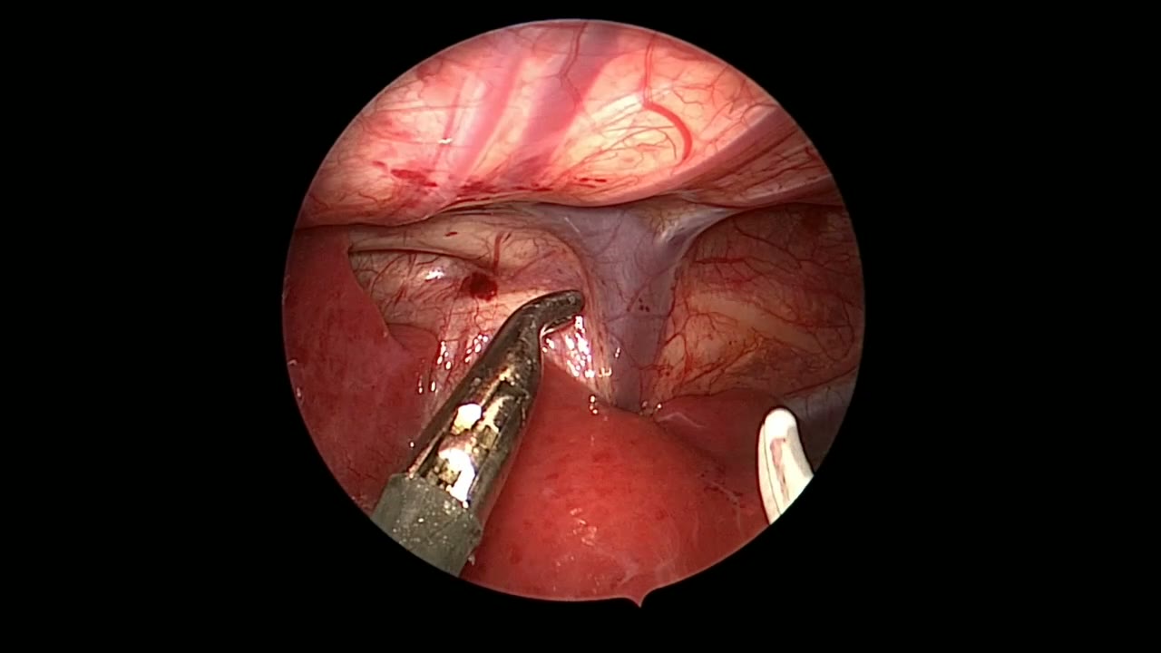
JRS TEF SHORT
Dr. Steve Rothenberg · 2 min · Published May 2026
Video
QUAD #10 Pt.3: Advantages of Thoracoscopic Repair of Esophageal Atresia with Dr. Steve Rothenberg
Dr. Steve Rothenberg · Published Jun 2024
Video
Intestinal Atresia Types Explained: Grossfeld Classification for Pediatric Surgery
1 min · Published Sep 2026
Video
Derivation and validation of the Pediatric Community-Acquired Pneumonia Severity (PedCAPS) score: A prospective cohort study
50 s · Published Sep 2026
Video
FETO for Late-Diagnosed Severe Congenital Diaphragmatic Hernia (CDH) at Cincinnati Children's with Dr. Beth Rymeski
4 min · Published Sep 2026
Video
Severe Congenital Diaphragmatic Hernia (CDH) Case: FETO Management & Unexpected Findings with Dr. Beth Rymeski
3 min · Published Sep 2026
Video
Fetoscopic Endoluminal Tracheal Occlusion (FETO) Procedure: Step-by-Step Guide with Dr. Beth Rymeski
4 min · Published Sep 2026
Video
Choledochal Cyst Types Explained: Pediatric Surgery Fundamentals
1 min · Published Sep 2026
What the experts said
The patient is a term male newborn with esophageal atresia and distal tracheoesophageal fistula.
Patient positioning is left lateral decubitus with the arm extended and restrained, with a roll under the left axilla.
Division of the azygous vein typically leads the surgeon to the fistula site.
The azygous vein can be divided with an energy device, simply coagulated, or double clipped and divided.
Thoracoscopic magnification allows for excellent delineation of the trachea and esophagus.
The fistula is ligated with a suture ligature flush with the trachea but not divided initially to prevent the distal esophagus from retracting.
Asking the anesthesiologist to push on the nasoesophageal tube aids in dissection of the proximal esophageal stump.
The esophagus is often quite adherent to the trachea and may even share a common wall, requiring careful dissection.
Dissection can be continued to the thoracic inlet, resulting in excellent mobilization of the proximal esophagus.
The fistula is divided a few millimeters distal to the ligature to liberate the distal esophagus.
Following fistula division, the ligature should be tested with 25 to 30 centimeters water positive pressure ventilation.
The back wall anastomosis is completed first using interrupted 4-0 or 5-0 absorbable sutures.
Extracorporeal knot tying was used in this case because the two ends came together nicely with minimal tension.
Sutures are placed 2 to 3 millimeters apart, with 5 to 6 sutures usually sufficient to complete the posterior wall anastomosis.
The nasoesophageal tube is advanced to protect the posterior wall prior to starting the anterior wall anastomosis.
It is important to take full thickness bites of healthy esophageal wall and mucosa with each suture.
Fewer sutures are usually required for the anterior wall anastomosis compared to the posterior wall.
A chest tube was placed in this case, but this is no longer routinely used.