


Chapter 1 of 5 · Surgical Management
Port placement
Patient positioning, port placement, and initial dissection of inferior pulmonary ligament
Expert statements on this page
No expert statements were drawn from this page.
Host summaries · secondary, not cited in answers
Educational content from recorded physician discussions — not medical advice. Talk to your (or your child's) care team about your situation.
Video
Thoracoscopic Left Lower Lobectomy for Congenital Pulmonary Airway Malformation
9 min · Published Feb 2020
Video
Clinical Symptoms Affect Treatment and Prognosis in Pediatric Patients with Congenital Pulmonary Airway Malformation
55 s · Published Dec 2023
Video
LLL intra lobar JRS
Dr. Steve Rothenberg · 4 min · Published May 2026
Video
Congenital Lung Malformations: CPAM, BPS, CLE, and Bronchogenic Cyst Explained
3 min · Published Oct 2026
Video
Thoracoscopic Right Lower Lobe Cystic Lesion Excision: Update Course 2014
Dr. Steve Rothenberg · 11 min · Published Sep 2014
Video
Quick Literature Updates Episode 18
4 min · Published Mar 2025
Video
sealer demo
Dr. Steve Rothenberg · 3 min · Published May 2026
Video
pyloric knife
Dr. Steve Rothenberg · Published May 2026
Video
pulm artery LLL
Dr. Steve Rothenberg · 9 s · Published May 2026
Video
plication narrated
Dr. Steve Rothenberg · 6 min · Published May 2026
Video
percuvance redo nissen
Dr. Steve Rothenberg · 5 min · Published May 2026
Video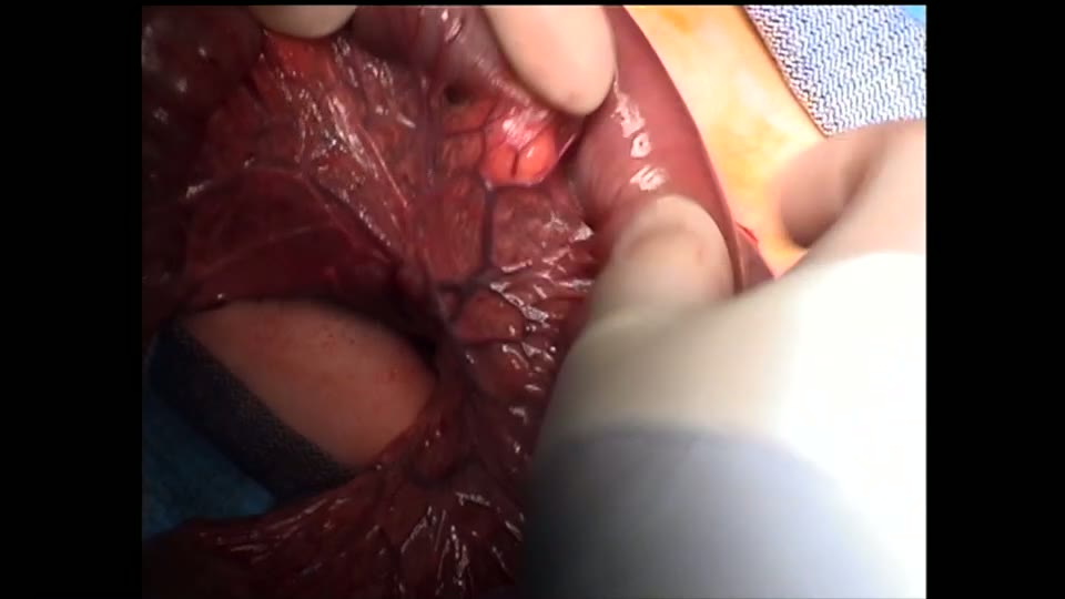
open bowel anast
Dr. Steve Rothenberg · Published May 2026
Video
Intestinal Atresia Types Explained: Grossfeld Classification for Pediatric Surgery
1 min · Published Sep 2026
Video
Derivation and validation of the Pediatric Community-Acquired Pneumonia Severity (PedCAPS) score: A prospective cohort study
50 s · Published Sep 2026
Video
FETO for Late-Diagnosed Severe Congenital Diaphragmatic Hernia (CDH) at Cincinnati Children's with Dr. Beth Rymeski
4 min · Published Sep 2026
Video
Severe Congenital Diaphragmatic Hernia (CDH) Case: FETO Management & Unexpected Findings with Dr. Beth Rymeski
3 min · Published Sep 2026
Video
Fetoscopic Endoluminal Tracheal Occlusion (FETO) Procedure: Step-by-Step Guide with Dr. Beth Rymeski
4 min · Published Sep 2026
Video
Choledochal Cyst Types Explained: Pediatric Surgery Fundamentals
1 min · Published Sep 2026
What the experts said
The patient is a 3-month-old who had a CPAM diagnosed prenatally.
The initial trochar is placed in the mid-axillary line in approximately the 6th intercostal space to overlook the major fissure.
The right and left hand operating ports are placed in the anterior axillary line in approximately the 5th and 8th intercostal spaces respectively.
The left-hand port is later changed to a 5 millimeter to accommodate the stapler.
The initial step is to take down the inferior pulmonary ligament and ensure there is not a systemic vessel or a hybrid lesion.
In this case, the fissure is incomplete anteriorly, although the vessels can be seen coursing through the fissure more posteriorly.
A 3 millimeter vessel sealer is used to seal the tissue and then divide sharply between it; this technique is both hemostatic as well as preventing air leaks in the divided tissue.
When the tissue is too thick, a sequential pattern is performed, sealing tissue anteriorly and then posteriorly, almost going through this as though doing a segmental resection in the liver.
Care must be taken to avoid injury to the apical or superior segmental branch, which usually comes off towards the posterior aspect of the fissure.
This vessel (apical segmental branch) usually is identified and separated and taken separately from the main trunk to the rest of the lower lobe.
Blunt dissection can be performed relatively aggressively once the anterior wall of the pulmonary vein is identified, as all tissue anterior to that can now be deemed safe for sealing and division.
Ideally, the seals will be 3 to 5 millimeters apart, and you can see the white bubble of the vessel between the two transparent seals; when seen like this, the seals are considered safe.
The vessel is divided partway to visualize the lumen, and once the lumen is seen and there is no bleeding, the vessel can be divided the rest of the way; this ensures that control of the vessel is not lost should there be a leak from either seal.
Even in this young child, there are already significantly enlarged lymph nodes which can obscure the dissection and make it hard to identify all the branches of the artery.
Depending on the size of the child and the size of the vessels, the basal segment can be taken as an entire trunk or can be done separately.
This child is a bit larger, almost 6 kg, and the vessels are taken after their branching point for safety; the smaller vessels are easier to seal and also provide some backup should there be a problem.
The 3 millimeter sealer has an upper limit of 5 millimeter vessel size that can be safely sealed.
If the distal seal causes disruption of the proximal seal because of the large size of the vessel and the fact that the seals are too close, the proximal seal is redone.
This technique of sealing proximally and distally to ensure good hemostasis and not allowing the vessels to retract has been used in over 500 thoracoscopic lobectomies with excellent results and without the occurrence of uncontrolled bleeding.
There is a pulmonary vein going to the apical or superior segment posteriorly, and care needs to be taken not to injure that during dissection of the apical segmental bronchus.
Prior to stapling, the bronchus is always crushed in order to help compress the tissue to enable the 5 millimeter stapler, which holds 2 millimeter staples.
The stapler is applied and closed for a count of 20, then fired, creating an excellent staple line with complete compression of the bronchus and complete closure.
In general, the bronchus to the basal segments is taken at the main takeoff and not further as was done with the vessels.
A 3 millimeter bowel clamp is inserted to crush the bronchus to the basal segments, as this tissue is thicker than the single segment taken before.
Care is taken not to cross over the previous staple line, as this can sometimes disrupt these smaller staples.
In general, the inferior pulmonary vein is taken as a solid trunk, but it is important to dissect high enough up on the vein to ensure that when applying the stapler, good proximal control can be obtained.
Occasionally it is necessary to take one or two small branches to gain adequate length on the trunk of the vein to apply the stapler.
If the vein branches more proximally, the speaker would simply dissect into the lung and get it at a segmental level using the sealer rather than the stapler.
It is critical that if the stapler is used on the inferior pulmonary vein trunk, there is proximal control in case there is a misfire, as this will allow further securing of the vein and eliminate the risk of uncontrolled bleeding.
The speaker has never had a problem using the stapler on the inferior pulmonary vein, but considers it imperative to take the precaution of proximal control, as if there were a problem, the vein could conceivably retract to the point where control could not be gotten thoracoscopically.