


From
StayCurrentMD
Lung Lesions: Fetal Interventions Parts I+II
Chapter 1 of 8 · Surgical Management
EXIT indications
EXIT procedure indications and ECMO use in CCAM
Expert statements on this page
No expert statements were drawn from this page.
Host summaries · secondary, not cited in answers
Educational content from recorded physician discussions — not medical advice. Talk to your (or your child's) care team about your situation.
Video
Fetal Interventions Part I: Lung Lesions
14 min · Published Aug 2012
Video
Fetal Interventions Part II: Lung Lesions
16 min · Published Jul 2017
Video
Prenatal Management of CPAMs: Lung Lesions
18 min · Published Aug 2012
Video
Open Fetal Surgery Overview: Fetal Surgery 2012
37 min · Published Jul 2015
Video
Postnatal Management of Lung Lesions Part I: Pediatric Thoracic Surgery Part...
Dr. Todd Ponsky · 31 min · Published Aug 2017
Podcast
The Full Story on CPAMs
56 min · Published Sep 2021
Video
Quick Literature Updates Episode 18
4 min · Published Mar 2025
Podcast
Journal of Pediatric Surgery Article Review: October 2023
12 min · Published Apr 2024
Video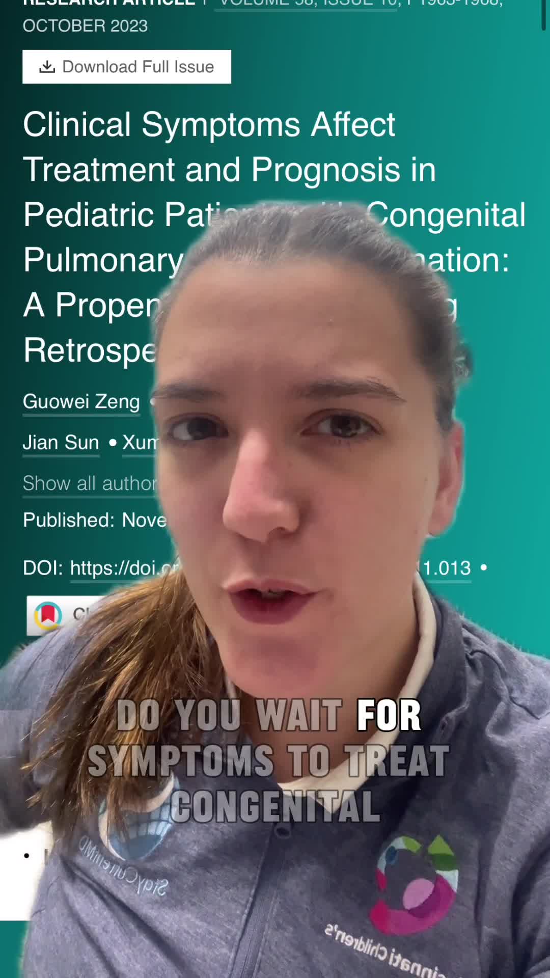
Clinical Symptoms Affect Treatment and Prognosis in Pediatric Patients with Congenital Pulmonary Airway Malformation
55 s · Published Dec 2023
Video
Update Course Rewind: Congenital Lung Lesions 2022
4 min · Published Aug 2023
Video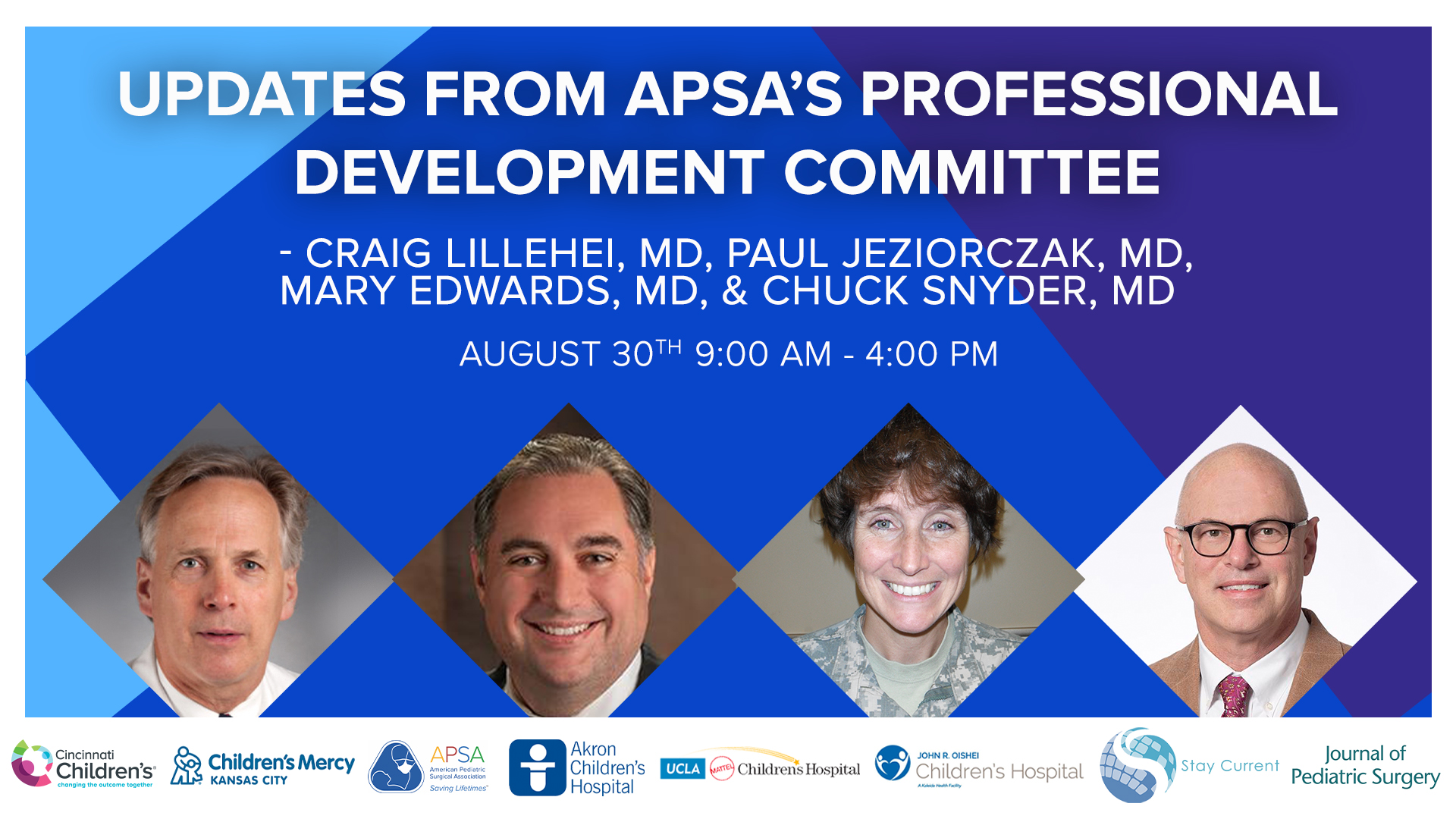
Update Course 2022 - APSA PDC UPDATES - Mary Edwards, Paul Jeziorczak, Craig Lillehei, and Charles Snyder,
64 min · Published Sep 2022
Podcast
Case Based Journal Review - CPAM in 2022
12 min · Published Apr 2022
Video
Intestinal Atresia Types Explained: Grossfeld Classification for Pediatric Surgery
1 min · Published Sep 2026
Video
Derivation and validation of the Pediatric Community-Acquired Pneumonia Severity (PedCAPS) score: A prospective cohort study
50 s · Published Sep 2026
Video
FETO for Late-Diagnosed Severe Congenital Diaphragmatic Hernia (CDH) at Cincinnati Children's with Dr. Beth Rymeski
4 min · Published Sep 2026
Video
Severe Congenital Diaphragmatic Hernia (CDH) Case: FETO Management & Unexpected Findings with Dr. Beth Rymeski
3 min · Published Sep 2026
Video
Fetoscopic Endoluminal Tracheal Occlusion (FETO) Procedure: Step-by-Step Guide with Dr. Beth Rymeski
4 min · Published Sep 2026
Video
Choledochal Cyst Types Explained: Pediatric Surgery Fundamentals
1 min · Published Sep 2026
What the experts said
EXIT procedures have been used for CCAMs with average CVR over 2, which are large lesions.
EXIT is reserved for cases with evidence of compression: diaphragmatic aversion, marked mediastinal shift, often ascites.
ECMO is very rare in CCAM patients and has not been used during EXIT procedures; conventional ventilation is tried first.
Referral centers receive complex cases while simple cases stay local, skewing the percentage requiring EXIT and fetal surgery.
There is no clear reduction in ability to have subsequent pregnancy after fetal surgery, based on approximately 4 published studies.
After fetal surgery hysterotomy (equivalent to classical cesarean in upper uterus), patients should never labor with future pregnancies.
No long-term maternal morbidity aside from requirement for cesarean delivery has been observed after fetal surgery.
Feared complications like placenta accreta at the hysterotomy site have not been observed thus far.
EXIT procedure requires an anesthetic team tuned into uterine relaxation and maternal issues, and expertise with hysterotomy.
EXIT procedure can be learned and disseminated more widely than fetal surgery without requiring a full fetal surgery program.
EXIT is valuable for cervical teratomas and airway obstructive problems; most large children's hospitals experience cases that would benefit from EXIT from time to time.
One center has performed close to 100 EXIT procedures in the past 10 years.
Maternal expertise must be available at the hospital; bringing a mother into a freestanding children's hospital without maternal expertise is a bad idea.
There are probably 4 or 5 centers in the US that clearly have the qualifications and background to do good EXIT procedures.
EXIT procedure involves weeks of planning, discussions of the entire team, and 15 to 20 people in the operating room.
If a center does only one EXIT per year, it is probably not enough to justify doing EXIT procedures; 3 to 5 per year is a reasonable threshold.
Very few BPS cases with large feeding vessels have been seen where high-output failure was thought to be the mechanism of hydrops; most have associated pleural effusions, mediastinal shift, or mass effect.
Injecting alcohol into fetal vessels has potential hazard including neurologic effects and has not been adequately studied.
Alcohol injection can cause thrombosis in systemic circulation; one case showed thrombi in heart chambers that then embolized to various sites.
Radiofrequency ablation cannot be controlled in the fetus due to 90% water content; energy can disperse unpredictably causing collateral damage.
In one experiment, radiofrequency probe placed in one side of a teratoma caused the other side to boil when activated.
Probably 95% of CCAMs seen now are prenatally diagnosed.
Almost none of prenatally diagnosed congenital lung lesions require prenatal intervention, and very few require intervention the day the child is born.
The vast majority of congenital lung lesions do not fall in the realm of extreme treatments; only one or two centers in the world should be thinking about these interventions.
The majority of children with prenatal lung lesions can be delivered without fetal distress.
Many lesions have been referred after a recommendation for termination by people who don't understand the natural history.
Even very large congenital lung lesions can regress and be asymptomatic at birth, or have very good survival rates with appropriate interventions.
The garden variety postnatal CCAM is very different than some prenatal CCAMs.
True cystic CCAMs do not disappear; they regress but remain present and prominent on CT scan one month after birth.
Some things that look like CCAMs in utero (like segmental bronchial stenosis) can give an echogenic microcystic appearance but may be minimally apparent or non-apparent after birth.
Congenital lung lesions should be called macrocystic, hyperechoic, or mixed rather than 'CCAM' prenatally, since CCAM is a pathological diagnosis once the specimen is in the bucket.
Many tertiary centers have the capacity to do EXIT; it requires a huge team approach and somebody leading the team.
Good prenatal diagnosis is essential; lung agenesis has been diagnosed as microcystic CCAM elsewhere, leading to potential catastrophic errors.
Pleuro-amniotic shunts are used specifically for macrocystic CCAMs with evidence of hydrops (pleural effusion, pericardial effusion, and skin or scalp edema), not pure ascites alone.
Large macrocystic lesions are sometimes tapped just prior to delivery to improve ventilation, but shunts are not placed prophylactically.
CVR less than 1.6 on presentation predicts about 3% (less than 5%) likelihood of evolving hydrops in microcystic lesions; this has held true in prospective studies and ongoing experience.
CVR greater than 1.6 requires close watching; there is much higher likelihood of evolving into hydrops.
Macrocystic lesions are a wild card because the cystic component can grow very rapidly and can be worrisome even if CVR is less than 1.6.
At one center, all lung lesions get a fetal MRI routinely; MRIs help define anatomy and general size of lesions.
MRIs are not essential for all centers; they can clarify abnormalities if there is ambiguity on ultrasound or confusion about diagnosis.