


From
StayCurrentMD
Spontaneous Pneumothorax: Update Course 2014
Chapter 1 of 4 · Diagnosis & Workup
Diagnostic pitfalls
Intraparenchymal disease mimicking effusion: diagnostic pitfalls
Expert statements on this page
No expert statements were drawn from this page.
Host summaries · secondary, not cited in answers
Educational content from recorded physician discussions — not medical advice. Talk to your (or your child's) care team about your situation.
Video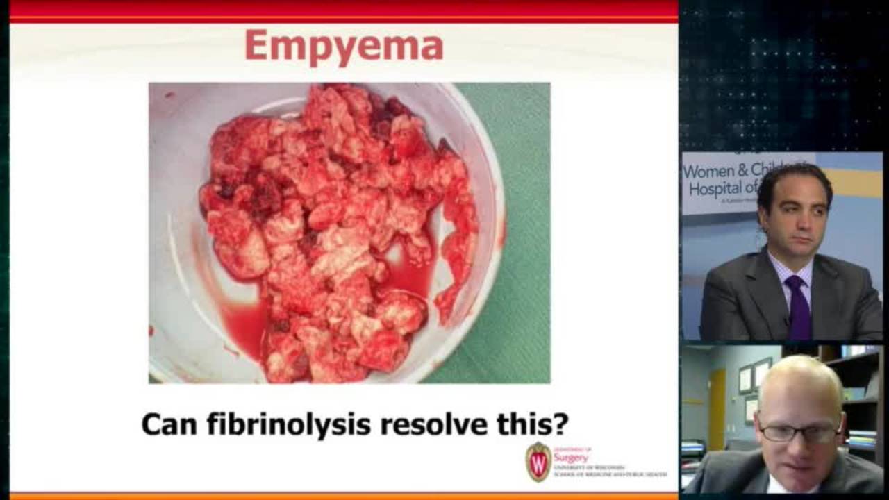
Empyema and Pneumothorax: Update Course 2014
31 min · Published Nov 2018
Podcast
Spontaneous Pneumothorax
33 min · Published Aug 2019
Video
Spontaneous Pneumothorax Rapid Fire: Update Course 2015
9 min · Published Nov 2015
Video
Supine Positioning for Bilateral VATS: Pediatric Surgery Difficult...
15 min · Published Jul 2017
Video
Tricks - Supine Positioning For Bilateral VATS
17 min · Published Nov 2018
Video
BOB Ped Surg 2023 - Jamie Schnuck, PAPS - Presentation
Published Feb 2023
Video
Intestinal Atresia Types Explained: Grossfeld Classification for Pediatric Surgery
1 min · Published Sep 2026
Video
Derivation and validation of the Pediatric Community-Acquired Pneumonia Severity (PedCAPS) score: A prospective cohort study
50 s · Published Sep 2026
Video
FETO for Late-Diagnosed Severe Congenital Diaphragmatic Hernia (CDH) at Cincinnati Children's with Dr. Beth Rymeski
4 min · Published Sep 2026
Video
Severe Congenital Diaphragmatic Hernia (CDH) Case: FETO Management & Unexpected Findings with Dr. Beth Rymeski
3 min · Published Sep 2026
Video
Fetoscopic Endoluminal Tracheal Occlusion (FETO) Procedure: Step-by-Step Guide with Dr. Beth Rymeski
4 min · Published Sep 2026
Video
Choledochal Cyst Types Explained: Pediatric Surgery Fundamentals
1 min · Published Sep 2026
What the experts said
Ultrasound can sometimes be misleading in distinguishing pleural effusion from intraparenchymal disease, requiring trust in ultrasound technician skill and correlation with CT when findings are discordant.
Intraparenchymal necrotic lung disease may require surgery a week or two later when the lung falls apart and develops an effusion and fistula.
For a 5-10% apical pneumothorax in a minimally symptomatic patient, observation with oxygen is appropriate initial management.
There is debate about whether oxygen therapy for pneumothorax actually works, though it is commonly used.
Simple aspiration of small pneumothorax may cause more trouble and result in a bigger pneumothorax.
Many pneumothorax patients never show an air leak after chest tube placement.
If simple aspiration is performed, leaving a catheter overnight may prevent the need for two procedures in patients who fail aspiration.
Needle aspiration of a small apical pneumothorax may result in a larger pneumothorax.
Simple aspiration is not well studied in children under 16 years old due to the rarity of spontaneous pneumothorax in this age group.
Patients with spontaneous pneumothorax typically have a very asthenic, thin chest configuration on X-ray.
Every spontaneous pneumothorax patient has blebs in the upper part of the lung.
CT is obtained to evaluate the contralateral side for blebs, which informs counseling about future risk and may guide decision for prophylactic intervention.
If CT shows contralateral blebs, a future pneumothorax on that side can be treated directly with VATS without wasting time on chest tube placement.
Before the era of VATS, the rule was to wait for a second pneumothorax before performing thoracotomy for bleb resection and pleural abrasion.
In the current era, the paradigm has shifted to more aggressive intervention with primary VATS on first pneumothorax presentation.
Prophylactic contralateral intervention may be justified in select cases such as patients planning extended remote travel (e.g., 2 months hiking in backcountry).
Standard VATS technique for pneumothorax includes apical wedge resection and roughing up the pleura.
If the patient has had multiple chest tubes previously, talc pleurodesis should be considered in addition to bleb resection and pleural abrasion.
Talc applied thoracoscopically distributes beautifully throughout the pleural space.
Mechanical abrasion of both visceral and parietal pleura improves outcomes, though aggressive visceral pleural abrasion can cause air leaks.
On re-operation, chests that had previous pleural roughing may appear as if no prior surgery was performed.
If all visible blebs are resected, pleurodesis may not be necessary.
Mechanical pleurodesis should be limited to the apex (4th intercostal space and above) rather than the entire chest.
Adequate mechanical pleurodesis is achieved when small blood vessels become prominent on the pleura.
Deflating and reinflating the lung multiple times during surgery helps identify blebs, particularly on the edges of the lower lobe.
Blebs can be sealed with energy devices rather than stapled, allowing surgery through 3 or 5 millimeter incisions.
A study of 350 patients who underwent lung procedures without postoperative chest tube placement showed no complications.
The no-chest-tube study included lung biopsies, not spontaneous pneumothorax patients.
Omitting chest tubes after pneumothorax surgery requires reliable nursing care to detect early postoperative pneumothorax, which may not be available in all settings.
Re-entering a chest that has had talc pleurodesis is extremely difficult and may require pleural decortication.
There is a 50% chance of failure with chest tube alone for spontaneous pneumothorax.
85% of pneumothorax patients can avoid an operation, making primary VATS potentially unjustified for all first-time presentations.
2001 ACCP consensus guidelines stated there was no role for simple aspiration in pneumothorax.
British Thoracic Society 2010 guidelines recommend simple aspiration as first-line therapy for pneumothorax.
A Cochrane review in 2007 found only one randomized controlled trial comparing manual aspiration versus chest tube for pneumothorax, which included patients as young as 16 years old.
The randomized trial showed an immediate success rate of aspiration of 59%, similar to chest tube success rates.
In the aspiration trial, there were 11 failures: 9 received chest tubes and only 2 went on to VATS, with 50% avoiding hospitalization and no difference in overall hospital stay or recurrence rate.
Every patient with spontaneous pneumothorax who has been evaluated in adult thoracic surgery has blebs on the top of the lung.
In adult thoracic surgery practice, the paradigm is to fix the symptomatic side with blebs and observe the contralateral side with blebs, intervening only if a second pneumothorax develops.
TissueSeal applied after roughing up pleural edges may promote adhesion, though it is more expensive than other methods.