


From
StayCurrentMD
Fetoscopic endoluminal tracheal occlusion and twin-twin transfusion: Fetal...
With Dr. Jan Deprest & Dr. Alan Flake & Dr. Michael Harrison & Dr. Eduardo Gratacos · hosted by Dr. Todd Ponsky
Chapter 1 of 6 · Fundamentals
FETO technique
Introduction and FETO Technique for Congenital Diaphragmatic Hernia
Expert statements on this page
No expert statements were drawn from this page.
Host summaries · secondary, not cited in answers
Educational content from recorded physician discussions — not medical advice. Talk to your (or your child's) care team about your situation.
Video
Fetoscopic Endoluminal Tracheal Occlusion (FETO) Procedure: Step-by-Step Guide with Dr. Beth Rymeski
4 min · Published Sep 2026
Video
Inside a FETO Procedure: Fetoscopic Balloon Tracheal Occlusion with Dr. Beth Rymeski
4 min · Published Jul 2026
Video
Tracheomalacia and tracheomegaly in infants and children with congenital diaphragmatic hernia managed with and without fetoscopic endoluminal tracheal occlusion (FETO): a multicentre, retrospective cohort study
59 s · Published Oct 2024
Video
Update Course 2021: TOP PUBLICATIONS IN NON - PED SURG JOURNALS
22 min · Published May 2022
Video
Yuichiro Miyake, MD - Best of the Best in Pediatric Surgery 2024
Dr. Todd Ponsky · 7 min · Published Feb 2024
Video
Controversies in Congenital Diaphragmatic Hernia: Update Course 2018
41 min · Published Aug 2018
Video
Utility of Fluorescence In Situ Hybridization as a Fetal Surgery Eligibility Criterion for....
52 s · Published Jun 2026
Video
The fetal frontier: A review of current and emerging fetal therapies for genetic diseases
44 s · Published May 2026
Video
Myelomeningoceles (open spina bifida) -Fetoscopic Intrauterine Myelomeningocele Closure
Lurie Children's Hospital · 3 min · Published Dec 2025
Video
Quick Literature Updates Ep 19
4 min · Published May 2025
Video
Open Fetal Surgery & EXIT Procedure with Dr. Jose Peiro
CCHMC Pediatric Surgery · 10 min · Published Oct 2024
Video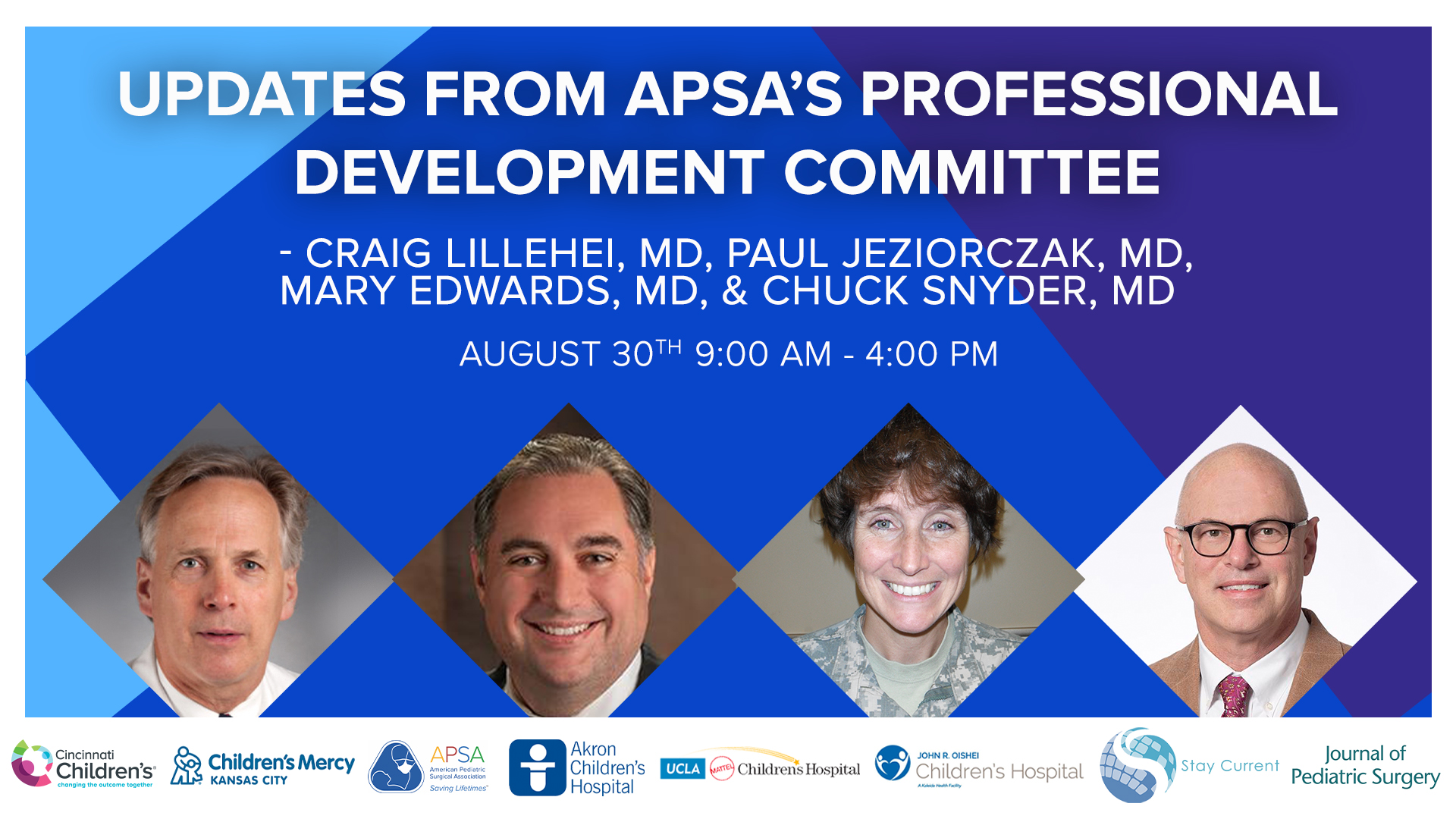
Update Course 2022 - APSA PDC UPDATES - Mary Edwards, Paul Jeziorczak, Craig Lillehei, and Charles Snyder,
64 min · Published Sep 2022
Only a few other public items share this expert — go deeper there →
Video
Intestinal Atresia Types Explained: Grossfeld Classification for Pediatric Surgery
1 min · Published Sep 2026
Video
Derivation and validation of the Pediatric Community-Acquired Pneumonia Severity (PedCAPS) score: A prospective cohort study
50 s · Published Sep 2026
Video
FETO for Late-Diagnosed Severe Congenital Diaphragmatic Hernia (CDH) at Cincinnati Children's with Dr. Beth Rymeski
4 min · Published Sep 2026
Video
Severe Congenital Diaphragmatic Hernia (CDH) Case: FETO Management & Unexpected Findings with Dr. Beth Rymeski
3 min · Published Sep 2026
Video
Choledochal Cyst Types Explained: Pediatric Surgery Fundamentals
1 min · Published Sep 2026
Video
Automated control of inspired oxygen in mechanically ventilated trauma and acute care surgery patients: A randomized controlled trial
48 s · Published Sep 2026
What the experts said
Congenital diaphragmatic hernia occurs in 10-20% of monochorionic twin pregnancies and is responsible for large amounts of perinatal mortality and morbidity, with 80-100% mortality if untreated in the past.
Patient selection for FETO is based on lung-to-head ratio (LHR) corrected for gestational age (observed/expected) and presence of liver herniation, with severe hypoplasia defined as maximum 20% survival, moderate 50%, and mild 85% survival.
The FETO procedure is done at 26-28 weeks (now 29 weeks in updated protocol) under local anesthesia with fetal analgesia and immobilization, lasting 6-10 minutes on average when fetal position is favorable.
The median gestational age at birth after FETO was 35 weeks, similar to open fetal surgery, with 20-22% experiencing ruptured membranes by 34 weeks.
Balloon removal in utero at least 24 hours prior to birth is associated with better survival and early morbidity compared to removal at delivery.
FETO increased survival from around 0% to 20% in the smallest lung group and by 30-35% on average in the severe hypoplasia group compared to historical controls.
Independent predictors of survival after FETO are initial lung size prior to operation, gestational age at delivery, and ability to remove balloon in utero at least 24 hours before birth.
Even with delivery prior to 32 weeks after FETO, survival rate is as high as predicted at term without fetal therapy; for delivery at 32 weeks or later, survival is 60%, which doesn't increase beyond 34 weeks.
There is an apparent decrease in bronchopulmonary dysplasia after FETO, with no evidence of substituting mortality by morbidity based on oxygen need, ventilator days, time to full enteral feeding, and NICU days.
The TOTAL trial in Europe compares tracheal occlusion at 27-29 weeks to expectant management, with survival as primary outcome in severe cases and survival without BPD in moderate cases.
Centers offering FETO must have an active fetoscopy program (minimum 36 interventions per year or 3 per month), experience with 15 balloon occlusion cases (at least 5 done locally), and 24-hour balloon removal capability.
In the moderate TTTS trial, 54 cases have been recruited beyond the first interim analysis, with all participating centers agreeing not to offer fetal therapy outside the trial.
The severe FETO trial has been hampered by too long offering the procedure outside trial settings while trying to optimize technique, though the last 100 cases showed no improvement in outcomes.
Ruben Quintero's 1997 staging system for TTTS actually describes physiologic states: stage 1 reflects initial volume transfer, stage 2 represents cephalization of blood flow, stage 3 represents increasing placental resistance, and stage 4 represents cardiac failure in the recipient.
Only 30-35% of stage 1 TTTS progresses to stage 2 or higher; 28% stay at stage 1 throughout pregnancy, and 40% regress completely or resolve.
The recipient twin in TTTS develops progressive hypertrophic cardiomyopathy due to both preload (volume) and afterload (peripheral vasoconstriction from renin-angiotensin activation) issues.
CHOP's cardiovascular score significantly drops within 1 week to 10 days after laser photocoagulation in the vast majority of cases, showing the recipient twin's heart has great potential to heal.
Diode laser is much safer than argon laser with lower penetration depth and no vessel rupture cases since switching to diode at CHOP.
CHOP's miss rate for vascular anastomoses is 0.8% based on placental injection studies, compared to 10-15% miss rates reported in literature.
CHOP has performed about 80 bipolar cord cauterizations with 86% singleton survival and over 90 radiofrequency ablations with 83% singleton survival for selective cord occlusion.
In Type 3 sIUGR, after laser photocoagulation to separate circulations, the smaller twin dies in 75% of cases within 48 hours because it was surviving on blood from the normal twin.
External validation from Children's Hospital Philadelphia (2006-2010 cohort) and Toronto confirmed that outcomes remain dependent on lung-to-head ratio and these selection criteria are still valid.
Amnio-reduction in severe polyhydramnios improves fetal hypoxia and reverses acidemia by decreasing amniotic fluid pressure, which reduces placental compression and improves blood flow.
Huber's 2006 study of over 200 laser cases showed 84% survival of at least one twin, 60% both-twin survival, and average delivery at 34 weeks, compared to 29 weeks with amnio-reduction.
High-volume laser centers achieve 93-94% survival of at least one twin, 88% overall survival, 78% both-twin survival, with average delivery at 34 weeks.
Major and minor neurologic deficits occur in 55% after amnio-reduction versus less than half that rate after laser, with most recent studies showing 5-6% major neurodevelopmental delays and 7-8% minor delays after laser.
Eduardo Gratacos classified selective IUGR into three types: Type 1 with positive end-diastolic flow (good prognosis), Type 2 with persistent absent/reversed flow (progressive deterioration), and Type 3 with intermittent cycling (unpredictable with high IUFD and 20% PVL risk in normal twin).
Type 3 sIUGR fetuses have the smallest placental share and characteristic very large arterio-arterial anastomoses that cause episodic acute fetal-to-fetal transfusions.
With purely expectant management of Type 2 sIUGR, there is 48% loss in the smaller twin and 33% in the normal twin, with only 37% intact survival in the smaller twin.
Type 3 sIUGR with expectant management shows 38.5% intact survival in the normal twin and 60% in surviving smaller twins, with very high morbidity and mortality in both groups.