


From
StayCurrentMD
Fetoscopic Endoluminal Tracheal Occlusion (FETO)
With Dr. Fung Lim · hosted by Dr. Rod Gerardo
Chapter 1 of 5 · Fundamentals
Introduction
Introduction to fetal surgery and congenital diaphragmatic hernia
Expert statements on this page
No expert statements were drawn from this page.
Host summaries · secondary, not cited in answers
Educational content from recorded physician discussions — not medical advice. Talk to your (or your child's) care team about your situation.
Video
Yuichiro Miyake, MD - Best of the Best in Pediatric Surgery 2024
Dr. Todd Ponsky · 7 min · Published Feb 2024
Video
Inside a FETO Procedure: Fetoscopic Balloon Tracheal Occlusion with Dr. Beth Rymeski
4 min · Published Jul 2026
Video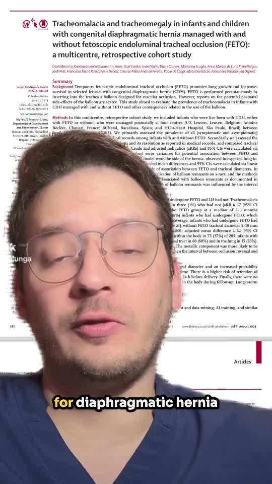
Tracheomalacia and tracheomegaly in infants and children with congenital diaphragmatic hernia managed with and without fetoscopic endoluminal tracheal occlusion (FETO): a multicentre, retrospective cohort study
59 s · Published Oct 2024
Video
Fetoscopic Endoluminal Tracheal Occlusion (FETO) Procedure: Step-by-Step Guide with Dr. Beth Rymeski
4 min · Published Sep 2026
Video
Quick Literature Updates Ep 19
4 min · Published May 2025
Video
Severe Congenital Diaphragmatic Hernia (CDH) Case: FETO Management & Unexpected Findings with Dr. Beth Rymeski
3 min · Published Sep 2026
Video
Utility of Fluorescence In Situ Hybridization as a Fetal Surgery Eligibility Criterion for....
52 s · Published Jun 2026
Video
The fetal frontier: A review of current and emerging fetal therapies for genetic diseases
44 s · Published May 2026
Video
Myelomeningoceles (open spina bifida) -Fetoscopic Intrauterine Myelomeningocele Closure
Lurie Children's Hospital · 3 min · Published Dec 2025
Video
Open Fetal Surgery & EXIT Procedure with Dr. Jose Peiro
CCHMC Pediatric Surgery · 10 min · Published Oct 2024
Video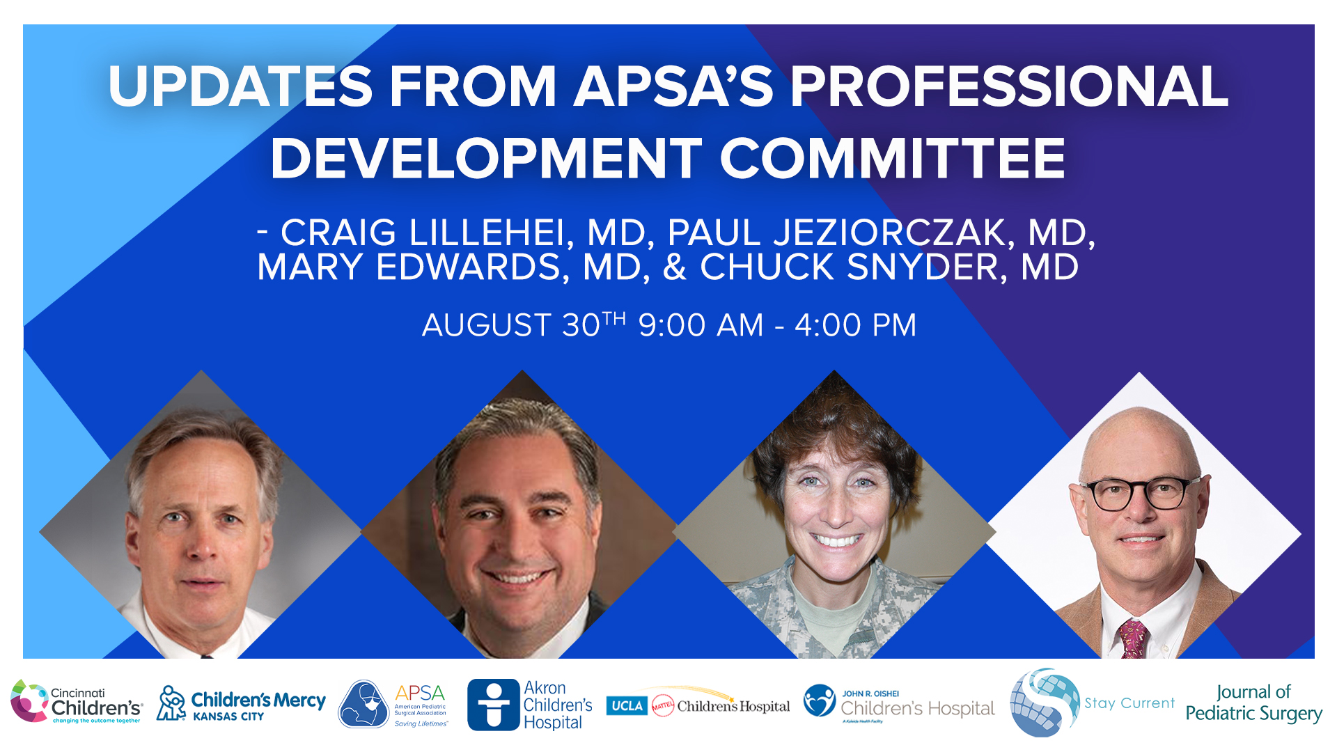
Update Course 2022 - APSA PDC UPDATES - Mary Edwards, Paul Jeziorczak, Craig Lillehei, and Charles Snyder,
64 min · Published Sep 2022
Podcast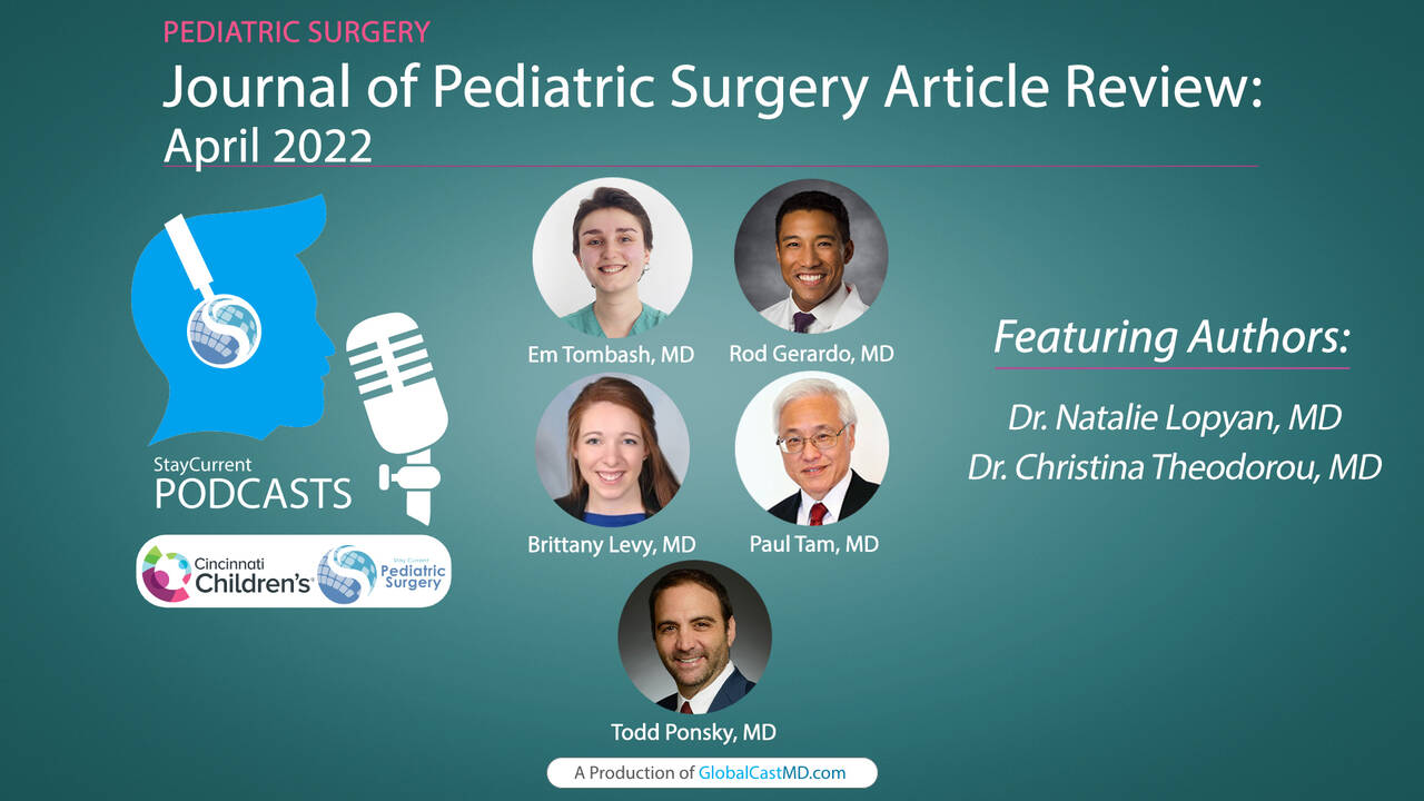
Journal of Pediatric Surgery Article Highlights: April 2022
8 min · Published Jul 2022
Video
Intestinal Atresia Types Explained: Grossfeld Classification for Pediatric Surgery
1 min · Published Sep 2026
Video
Derivation and validation of the Pediatric Community-Acquired Pneumonia Severity (PedCAPS) score: A prospective cohort study
50 s · Published Sep 2026
Video
FETO for Late-Diagnosed Severe Congenital Diaphragmatic Hernia (CDH) at Cincinnati Children's with Dr. Beth Rymeski
4 min · Published Sep 2026
Video
Choledochal Cyst Types Explained: Pediatric Surgery Fundamentals
1 min · Published Sep 2026
Video
Automated control of inspired oxygen in mechanically ventilated trauma and acute care surgery patients: A randomized controlled trial
48 s · Published Sep 2026
Video
Esophageal Atresia & Tracheoesophageal Fistula (EA/TEF) Types Explained for Pediatric Surgery
2 min · Published Sep 2026
What the experts said
It is difficult to determine with imaging exactly how large the hole in the diaphragm is in CDH.
In mild diaphragmatic hernia, the left lung starts to shrink in size.
In moderate diaphragmatic hernia, the left lung gets smaller as the intestines and part of the liver push upwards.
In the most severe CDH cases, the liver occupies a good portion of the chest, the left lung is very small, and even the right lung is shrinking.
The tracheal occlusion procedure is commonly performed at gestational age between 27 weeks and 29 weeks 6 days.
Anesthesia for FETO is induced by ultrasound guidance with local anesthetic and numbing medication injected into the mother.
An introducer is inserted into the amniotic space to allow placement of a fetoscope (small camera) through the introducer into the amniotic space.
The fetoscope is advanced carefully into the fetal trachea once the baby's mouth is located.
The ideal position for the fetoscope in FETO is in the main trachea below the vocal cords but above the carina, before the trachea splits into the two main bronchi.
A balloon is inserted into the fetal airway, inflated to completely occlude the trachea, then detached and left in place.
The balloon is left in place for a few weeks to accelerate lung growth.
If the baby is in proper position, the balloon can be punctured under ultrasound guidance.
The deflated balloon is pushed out of the baby's trachea by lung fluids and poses no risk to the baby's health.
If the baby's position does not allow for needle puncture, a grasper is used to hold the balloon while a needle punctures it, and the deflated balloon is then removed from the airway using the grasper.
After balloon removal, the mother and fetus are monitored carefully for the remainder of the pregnancy.
Ideally, the baby is delivered vaginally at term, with cesarean section reserved for the usual obstetrical reasons.
Ultrasound is usually the screening tool to find congenital diaphragmatic hernia in the fetus.
MRI provides higher resolution imaging than ultrasound and yields information about the pulmonary status of the fetus in CDH.
Fetuses with severe CDH (liver herniation and bilateral lung hypoplasia) are good candidates for FETO.
Fetal lung tissue constantly creates fluid that normally escapes through the trachea; occluding the trachea causes fluid and pressure to build up, which helps the lungs develop.
The balloon is typically removed at about 34 weeks gestation.