


From
Dr. Marc Levitt
Colorectal Quiz Episode 17: Cloaca Part 1
With Dr. Marc Levitt & Dr. Richard Wood · hosted by Dr. Amanda Jensen & Dr. Kira Ahmad
Chapter 1 of 7 · Fundamentals
Case presentation
Introduction and Case Presentation: 31-Week Infant with Single Perineal Orifice
Expert statements on this page
No expert statements were drawn from this page.
Host summaries · secondary, not cited in answers
Educational content from recorded physician discussions — not medical advice. Talk to your (or your child's) care team about your situation.
Video
Cloaca - Workup & Evaluation
20 min · Published Nov 2018
Video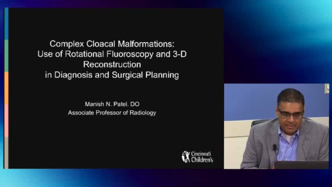
3D Reconstruction: Cloaca and Complex ARMs 2015
CCHMC Pediatric Surgery · 20 min · Published Oct 2015
Podcast
Cloaca Management with Dr. Marc Levitt & Dr. Aaron Garrison
43 min · Published Dec 2016
Video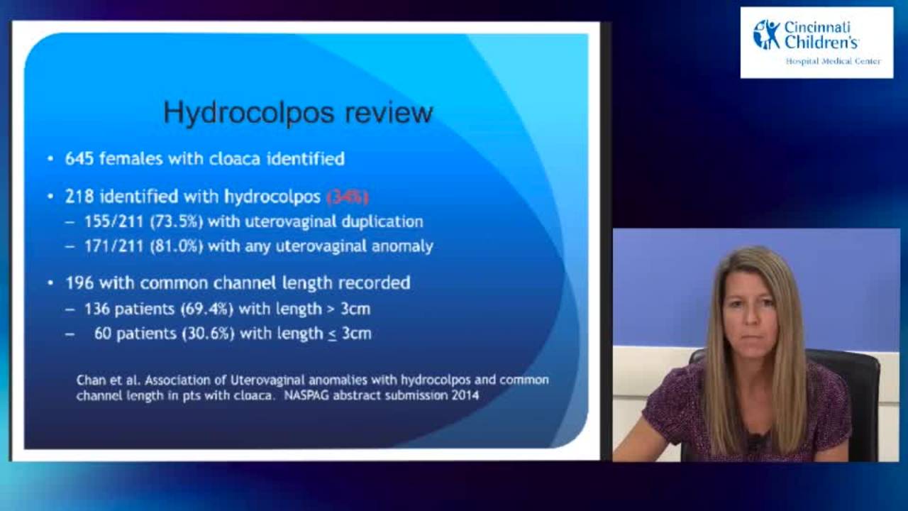
Case Presentations Part I: Cloaca and Complex ARMs 2015
45 min · Published Oct 2015
Video
Cloaca - Case Presentations
45 min · Published Nov 2018
Video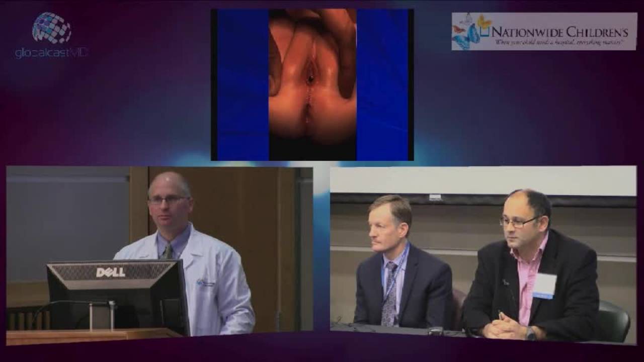
Anorectal Malformation Management of Female Patients Part II: Pediatric...
28 min · Published Apr 2012
Video
Validation of an anorectal malformation trainer - Can a high-fidelity model simulate real life?
1 min · Published Jun 2026
Video
Complications and Long-Term Outcomes of Patients With Cloacal Malformation After Bowel Neovagina...
1 min · Published Jun 2026
Video
Safety and utility of long-acting steroid injection for management of post-operative stricture...
1 min · Published Jun 2026
Video
Association Between Social Determinants of Health and Choice of Urinary Reconstruction in Children
1 min · Published Jun 2026
Video
The Perineal Body Preserving PSARP (PPP)
11 min · Published Jun 2026
Video
Update Course Rewind 2025: Hirschsprung + ARM: Rare but Real
1 min · Published May 2026
Video
Colorectal Quiz Ep. 50 -16th Annual European Pediatric Colorectal & Pelvic Reconstruction Conference
Marc Levitt · Published Mar 2026
Podcast
Colorectal Quiz: Episode 50 - 16th Annual European Pediatric Colorectal and Pelvic Reconstruction Conference, Stockholm, Sweden, October 2025 - What did we learn?
24 min · Published Mar 2026
Video
Colorectal Quiz Episode 33: Cloaca Exstrophy
Marc Levitt · 22 min · Published Sep 2025
Video
The Colorectal Quiz Episode 21: The History of Hirschsprung Disease
Marc Levitt · 15 min · Published Sep 2025
Video
Colorectal Quiz: Episode 49 – Collaborating for Kids: Colorectal & Pelvic Care (with Help from AI)
Marc Levitt · 40 min · Published Jul 2025
Podcast
Colorectal Quiz: Episode 49 - Collaborating for Kids: Colorectal & Pelvic Solutions (with a Little Help from AI)
Marc Levitt · 40 min · Published Jul 2025
What the experts said
A single perineal orifice in a newborn indicates cloaca, where the vagina, urethra, and rectum are fused together inside creating a single common channel.
Hydrocolpos is the distension of the vagina caused by accumulation of fluid.
Cloaca or anorectal malformation is associated with VACTERL and needs to be worked up as such.
VACTERL association requires three or more anomalies: vertebral, imperforate anus, cardiovascular, tracheoesophageal fistula, esophageal atresia, renal/radial, and limb defects.
The diagnostic yield for cloaca on prenatal ultrasound is still much lower than desired.
Hydrocolpos on prenatal ultrasound should alert to the possibility of cloaca.
Prenatal findings suggestive of cloaca include abnormal kidneys, single kidney, and two-vessel cord association.
A smart perinatologist seeing a female fetus with pelvic mass and kidney abnormalities should consider cloaca diagnosis, which influences delivery location.
Neonatologists might incorrectly conclude cloaca is ambiguous genitalia and do unnecessary endocrine workup.
Good physical exam with good lighting is the first step in evaluating suspected cloaca.
On exam, distracting the labia reveals more of a clitoral hood than real labia minora, with a single perineal orifice posterior to the clitoral hood.
A perineal groove behind the labia structures and dimpling representing muscle complex suggests the perineum is not completely flat.
Children with cloacal malformation who do not have an anus do not need investigation for ambiguous genitalia or disorders of sexual differentiation because they are known to be female.
Initial urgent management priorities are ensuring kidney decompression/urine drainage, diagnosing hydrocolpos, and confirming safety for OR (ruling out TEF and cardiac issues).
Initial workup should consist of NG tube and chest x-ray, cardiac echo, and pelvic and renal ultrasound to assess for TEF, cardiac anomalies, hydrocolpos, and hydronephrosis.
Bilateral hydronephrosis with hydrocolpos requires management of the hydrocolpos as part of initial treatment.
The modern approach has moved heavily away from vaginostomy toward clean intermittent catheterization through the common channel for hydrocolpos drainage.
To perform CIC for hydrocolpos, pass a tube through the common channel, drain fluid, get ultrasound to confirm the tube is in the hydrocolpos/vagina, confirm decompression, then continue recurrently.
If CIC effectively drains the hydrocolpos, proceed with colostomy only and continue drainage postoperatively; if unsuccessful, can return to formal vaginostomy.
Seattle Children's (Paul McGarrian, Jeff Evan Sino, Caitlin Smith) demonstrated that many hydrocolpi can be drained perineally, changing the previous dogma of routine vaginostomy.
When catheterizing for hydrocolpos drainage, you are more likely to get into the vagina than the bladder due to the anatomy of the urethral takeoff to the bladder neck.
The modern hydrocolpos drainage approach is published in a Seminars in Pediatric Surgery article but not yet in textbooks.
To confirm proper catheter placement, pass the tube, leave it in, get bedside ultrasound within first 24 hours to confirm it's in the hydrocolpos and decompressing it.
During ultrasound-guided drainage, when you drain the hydrocolpos, the bladder immediately fills, demonstrating the physiology where hydrocolpos obstructs the ureters.
A vesicostomy is the wrong move in almost every cloaca; the hydrocolpos needs to be drained instead.
Once hydrocolpos is drained, ureters are no longer compressed at the trigone and can empty into the bladder, which then empties out the common channel or back into hydrocolpos for sequential drainage.
Catheterization frequency is typically three times daily initially, then twice daily when family takes over, with serial ultrasounds every 2-3 days initially, then weekly, then monthly after discharge.
The most important measure of successful hydrocolpos drainage is kidney decompression, not the hydrocolpos itself; if kidneys are completely normal, the hydrocolpos doesn't matter.
Even with vaginostomy tubes, you must continue checking that kidneys remain decompressed; don't assume the tube is doing its job without verification.
Newborn management bullet points: good exam with good light to diagnose, no endocrine workup for cloaca, renal/pelvic ultrasound plus tests for anesthesia safety, drain hydrocolpos by CIC, and colostomy within 24-48 hours.
Colostomy should be done as proximally as possible (descending-sigmoid junction) to ensure enough length for distal work, rather than lower sigmoid.
Laparoscopy for newborn colostomy in non-distended patients provides good pelvic anatomy visualization, allows precise colostomy site selection, and avoids wound between stomas.
Laparoscopic technique: mobilize lateral attachments of descending colon, bring bowel up through mucous fistula site, staple it, wash out distal limb completely, then make separate incision for proximal stoma with no surrounding incision for clean skin and easy bagging.
For vaginostomy in patients with vaginal septum, open the anterior wall of the hydrocolpos vagina and remove a small portion of septum to drain both sides through one hole.
Vaginostomy can be done with or without tubes; tubes can become encrusted and colonized, so tubeless has some advantage if anatomy allows easy reach to abdominal wall.
For massive hydrocolpos requiring open approach, use lower midline incision to get above the hydrocolpos, which is very adherent to anterior abdominal wall and inflamed; standard left lower quadrant incision will cause trouble.
For large hydrocolpos via midline: open into dome, remove bit of septum, close it, put in tube to drain both sides, or suture to abdominal wall like vesicostomy/gastrostomy to avoid indwelling tube as nidus for infection.
Critical distinction: single perineal orifice with no anal opening is cloaca (female, no endocrine workup); completely normal anus with perineal orifice is urogenital sinus (needs endocrine workup for CAH, electrolyte check).
Urogenital sinus patients can also have hydrocolpos and hydronephrosis with similar management, but no colostomy needed since they have an anus.
Post-discharge follow-up focuses on ensuring kidneys are well decompressed, patient is growing well, following kidney function tests, and ensuring parents manage stoma effectively.
With effective urine and stool drainage, patients should be thriving unless other underlying issues exist.
In the large majority of cloaca patients, diagnosis is made at birth rather than prenatally.
Definitive imaging and reconstructive planning should wait until the patient is growing and thriving.